
When we imagine the prehistoric world, the mind often conjures images of towering giants, primitive oceans, and a relentless struggle for survival. We see the strength of the apex predator and the speed of the prey, but we rarely consider the fragility that accompanied them. For a long time, the fossil record was viewed primarily as a ledger of species and extinctions—a map of who lived and when they vanished.
However, a growing field of study is revealing that the history of life is also a history of illness. From the depths of the Paleozoic seas to the terrestrial reign of the dinosaurs, disease has been a constant companion to biological existence. This intersection of paleontology and medicine, known as paleopathology, allows scientists to move beyond the anatomy of a species and begin reconstructing the actual lived experiences—and the suffering—of individual organisms.
By analyzing anomalous alterations in fossilized remains, researchers are discovering that the biological responses to injury and infection have remained remarkably consistent across millions of years. This scientific lens transforms a piece of stone into a medical chart, revealing that the “fragility” we associate with the human condition is, in fact, an ancient trait shared by nearly every complex organism that has ever walked or swam on Earth.
For those of us focused on the evolution of technology, the most fascinating aspect of this research is not just what is being found, but how. The leap from visual inspection of a bone to high-resolution internal imaging has revolutionized our understanding of prehistoric health, turning paleontology into a high-tech diagnostic exercise.
The Mechanics of Paleopathology: Bridging Past and Present
Paleopathology is the scientific discipline dedicated to the analysis of pathological alterations in fossil remains. Its primary goal is to identify and interpret the ailments that affected extinct organisms, providing critical data on their biology, ecology, and the evolutionary trajectory of diseases.

The discipline operates on a fundamental premise: biological processes respond in comparable ways across different species and throughout evolutionary time. To reach a diagnosis for a creature that has been dead for millions of years, scientists use a comparative method. They analyze a lesion or a deformity in a fossil and compare it to known pathologies in extant (living) species. If a bone growth in a dinosaur resembles the arthritis seen in modern birds or mammals, researchers can infer a similar degenerative process occurred in the prehistoric animal.
While pathological processes have been identified in a vast array of extinct organisms—ranging from simple protozoa to complex vertebrates—they are most frequently documented in groups with “hard parts.” Bones, shells, and exoskeletons are far more likely to fossilize than soft tissue, meaning our understanding of ancient disease is heavily weighted toward skeletal and structural ailments.
Digital Diagnostics: The Role of CT Scanning in Paleontology
The evolution of paleopathology has been inextricably linked to the evolution of medical imaging. In the past, diagnosing a fossil often required invasive techniques or relied solely on surface-level observations, which could be misleading. Today, the field has adopted the same tools used in modern hospitals to treat living patients.
High-resolution scanners and Computed Tomography (CT) scans are now central to the process. These tools allow paleontologists to observe internal structures and tissues without damaging the specimen. By creating a three-dimensional digital map of the fossil, researchers can detect bone infections, identify the specific way a fracture remodeled itself during healing, or spot developmental deformities that are invisible to the naked eye.
This non-destructive approach is critical because fossils are irreplaceable. The ability to “slice” through a fossil digitally means that scientists can conduct multiple analyses over decades as imaging software improves, without ever risking the physical integrity of the specimen. It is, a digital autopsy performed on a patient who has been dead for an eternity.
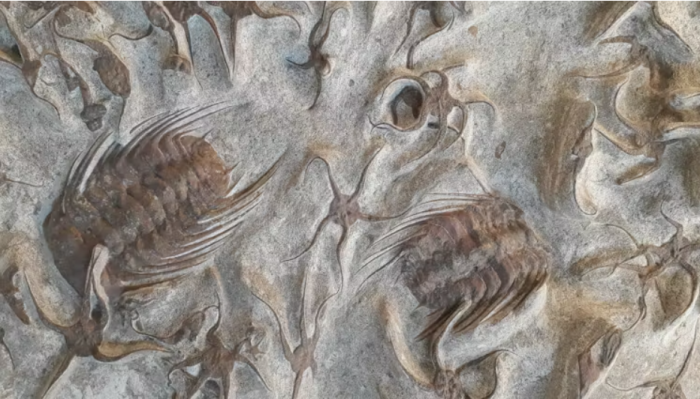
Survival in the Paleozoic: The Resilience of Trilobites
One of the most striking examples of ancient resilience is found in the trilobites. These marine arthropods were symbols of the Paleozoic era, with more than 22,000 described species that dominated the oceans for hundreds of millions of years. Because they possessed hard exoskeletons, they provide an excellent record for paleopathologists.
For years, paleontologists found trilobite fossils with truncated shells, deep notches, or missing sections. These were often dismissed as “taphonomic” damage—meaning the shell was simply broken after the animal died or during the fossilization process. However, closer inspection has revealed a different story.
Many of these specimens reveal evidence of regenerated edges. This indicates that the trilobites suffered brutal attacks from predators but survived. The presence of healed edges on a shell proves that the organism lived long enough for its biological repair mechanisms to kick in, offering a rare glimpse into the survival instincts and regenerative capabilities of early marine life.
Dinosaur Health: From Fractures to Arthritis
The dinosaur record is equally revealing, showing that the giants of the Mesozoic were susceptible to many of the same ailments that plague modern vertebrates. Their bones frequently bear the marks of a hard life, providing a narrative of injury and recovery.
Researchers have identified several recurring pathological patterns in dinosaur fossils:
- Healed Fractures: Many fossils show evidence of “soldered” fractures, where the bone has successfully fused back together. This suggests that some dinosaurs were able to survive significant trauma and recover, despite the lack of medical intervention.
- Degenerative Diseases: There is evidence of conditions similar to arthritis in dinosaur joints, suggesting that age and wear-and-tear affected their mobility just as it does in modern animals.
- Infections and Amputations: Some specimens show signs of chronic bone infections or the results of amputations, where a limb was lost but the animal continued to live for a period afterward.
These findings humanize these prehistoric creatures, shifting the narrative from indestructible monsters to biological entities that experienced pain, illness, and the slow process of healing.
Why Ancient Illness Matters Today
The study of paleopathology is more than a historical curiosity; it is a vital component of understanding the broader history of life on Earth. By documenting how diseases manifested in the past, scientists can better understand the origin, distribution, and evolution of pathogens and biological failures.
This research provides a baseline for evolutionary biology. When we see that degenerative joint diseases existed millions of years ago, it tells us that these conditions are not necessarily the result of modern lifestyles, but are inherent risks of having a skeletal structure. Similarly, seeing the regenerative success of trilobites helps scientists understand the evolution of healing mechanisms in arthropods.
these insights help ecologists reconstruct ancient environments. The prevalence of certain injuries or infections can indicate the types of predators present in an ecosystem or the nutritional stresses the population was facing.
Key Insights into Paleopathology
| Organism Group | Observed Pathologies | What it Reveals |
|---|---|---|
| Trilobites | Regenerated shell edges, deep notches | Survival of predator attacks and regenerative capacity. |
| Dinosaurs | Healed fractures, arthritis-like degeneration | Capacity for bone remodeling and age-related joint decay. |
| General Vertebrates | Bone infections, developmental deformities | Consistency of biological responses to infection over eons. |
As technology continues to advance, the resolution of our “digital autopsies” will only improve. We are moving toward a future where we might identify specific prehistoric pathogens or understand the cellular response to ancient traumas. The work of researchers like Blanca Moncunill Solé, a postdoctoral researcher at Universidade da Coruña, underscores the importance of this interdisciplinary approach, combining biology, geology, and advanced imaging to read the hidden medical histories of the Earth.
The next major checkpoint for the field will likely involve the integration of even higher-resolution synchrotron imaging, which promises to reveal cellular-level details in fossils that were previously thought to be unreachable. As these tools grow more accessible, our understanding of the “fragile” side of prehistory will only grow.
Do you think the ability to “diagnose” extinct species changes how we view the evolution of life? Share your thoughts in the comments below.