
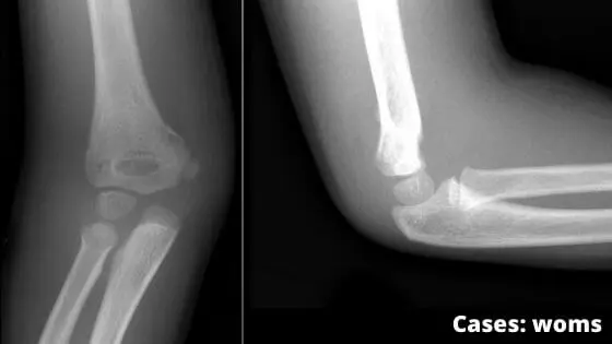
In the realm of orthopedic trauma, few injuries present as distinct a clinical challenge as the supracondylar fracture of the humerus. While this injury is most frequently associated with pediatric patients—often occurring in children aged 5 to 7 following a fall on an outstretched hand—it remains an important consideration in clinical practice due to its potential for long-term complications. As a physician, I have seen how these fractures, which occur at the distal end of the humerus just above the elbow joint, require precise diagnostic evaluation to ensure the neurovascular integrity of the arm is maintained.
When a patient presents with an acute elbow injury, the primary goal for any clinician is to assess the stability of the fracture and rule out damage to the nerves and blood vessels that traverse the elbow. While clinical observation is the first step, definitive management often relies on radiographic imaging to classify the fracture pattern. According to clinical guidance provided by the Royal Children’s Hospital Melbourne, these fractures are initially categorized based on the direction of displacement, which dictates whether a patient requires simple immobilization or more invasive surgical intervention.
Understanding the Fracture Mechanism and Clinical Presentation
A supracondylar fracture of the humerus occurs in the thin, distal portion of the bone, situated just above the medial and lateral condyles. In the pediatric population, the bone is still developing, making this region a common site for trauma. When a child falls, the force is transmitted through the forearm to the elbow, causing the distal fragment to shift. While the majority of these injuries are non-displaced and can be managed effectively with casting, those that involve significant angulation or displacement often necessitate surgical reduction to prevent complications such as malunion or restricted range of motion.
The symptoms are usually immediate and unmistakable: severe pain, pronounced swelling around the elbow and a significant loss of function in the affected limb. It is crucial for healthcare providers to monitor for late-onset pain, which can be an indicator of muscle ischemia—a condition where the muscles do not receive sufficient oxygen. If left unaddressed, this can lead to permanent loss of muscle function. Medical professionals perform a thorough neurovascular assessment, checking for distal pulses and testing the function of the radial, median, and ulnar nerves to ensure the injury has not compromised blood flow or nerve signaling to the hand.
Diagnostic Challenges and Neurovascular Assessment
One of the most critical aspects of diagnosing a supracondylar fracture is the evaluation of nerve integrity. The anterior interosseous nerve, a branch of the median nerve, is particularly vulnerable during postero-lateral displacement of the distal humerus. Clinicians often look for a weakened “OK” sign—where the patient is unable to form a perfect circle with their thumb and index finger—as a clinical indicator of nerve involvement. In such cases, a pincer grasp is often substituted by the patient during physical examination.
If pulses are not palpable, or if there is concern regarding the blood supply to the arm, Doppler ultrasonography is frequently employed to ascertain the status of the brachial artery. The goal of these diagnostic efforts is to prevent the onset of compartment syndrome, a serious complication where pressure within the muscles increases to dangerous levels. By identifying these issues early, orthopedic teams can act swiftly to restore alignment and ensure the limb remains viable.
Treatment Pathways and Long-Term Outlook
The prognosis for a supracondylar fracture is generally positive, provided the injury is managed correctly. In children, the body’s natural capacity for bone remodeling is often sufficient to achieve a full recovery after the fracture has been stabilized. However, the treatment plan is highly individualized based on the fracture’s severity. Non-displaced fractures typically involve protective casting, while displaced or angulated fractures require the surgeon to realign the bone fragments, often using pins under fluoroscopic guidance to hold the humerus in the correct position while it heals.

As we move forward in orthopedic medicine, the focus remains on minimizing the need for invasive procedures while maximizing functional outcomes. Ongoing research continues to refine the criteria for surgical intervention, helping surgeons decide exactly when a fracture is stable enough for conservative management and when it requires the precision of a surgical theater. For patients and parents, the most important takeaway is to seek immediate evaluation if an elbow injury follows a fall, as timely intervention is the single most effective way to avoid the long-term complications associated with this common yet complex injury.
As this remains a standard topic in pediatric orthopedics, patients and families are encouraged to consult their local healthcare providers for updates on regional treatment protocols and to share their experiences with recovery in the comments section below.