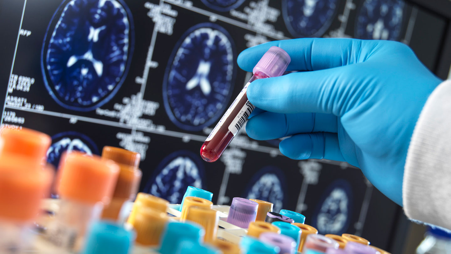
Researchers are developing an experimental blood test that uses artificial intelligence to identify overlapping types of dementia. By analyzing complex protein patterns, the test aims to distinguish between Alzheimer’s disease, Lewy body dementia, and other neurodegenerative conditions, potentially allowing for more precise treatment in patients with mixed pathologies.
Current diagnostic methods often struggle to identify patients who suffer from more than one form of cognitive decline simultaneously. While traditional tests frequently focus on a single disease marker, new research into blood-based proteomics suggests that machine learning can detect the distinct “signatures” of multiple diseases within a single blood sample. This development could move clinical practice away from broad diagnoses toward highly specific, personalized neurological care.
For decades, the gold standard for diagnosing neurodegenerative diseases has relied on expensive imaging or invasive procedures. However, as the global population ages, the demand for scalable, cost-effective screening tools has increased. The integration of AI into blood-based biomarker analysis represents a significant shift in how clinicians approach the complexities of the aging brain.
The clinical challenge of mixed dementia
In clinical practice, dementia is rarely a monolithic condition. Many patients present with “mixed dementia,” a state where multiple pathological processes occur in the brain at once. According to research published in various neurological journals, a significant portion of elderly patients exhibit features of both Alzheimer’s disease and vascular dementia, or Alzheimer’s combined with Lewy body dementia.
This overlap creates a diagnostic hurdle. Symptoms like memory loss, confusion, and motor impairment can be attributed to several different underlying causes. If a physician treats a patient for Alzheimer’s when the primary driver is actually Lewy body dementia, the treatment plan may be ineffective or even counterproductive. The inability to distinguish these overlapping conditions often leads to misdiagnosis or delayed intervention.
The complexity of these diseases stems from the different proteins involved in their progression. Alzheimer’s is characterized by the accumulation of amyloid-beta and tau proteins. In contrast, Lewy body dementia is primarily associated with the protein alpha-synuclein. Identifying which proteins are present, and in what concentrations, is essential for an accurate diagnosis. Traditional methods struggle to map these concurrent protein pathologies without high-cost intervention.
How AI-driven proteomics identifies disease signatures
The experimental approach relies on proteomics—the large-scale study of proteins. Instead of looking for a single biomarker, such as a specific level of p-tau217, researchers are using machine learning to scan hundreds or even thousands of proteins in the blood simultaneously. This method looks for “proteomic fingerprints” that characterize specific neurodegenerative states.
Artificial intelligence is uniquely suited for this task because the relationships between these proteins are non-linear and highly complex. A human clinician cannot manually calculate the interaction of 500 different protein concentrations to determine a diagnosis. However, machine learning models can be trained on large datasets of known disease profiles to recognize the subtle patterns that signal specific types of neurodegeneration.
By identifying these patterns, the AI can potentially flag the presence of multiple pathologies. For example, the model might detect a signature that contains both the amyloid-beta markers of Alzheimer’s and the alpha-synuclein markers of Lewy body dementia. This capability would allow doctors to confirm a mixed dementia diagnosis through a routine blood draw rather than a complex series of specialized scans.
The role of machine learning in biomarker discovery
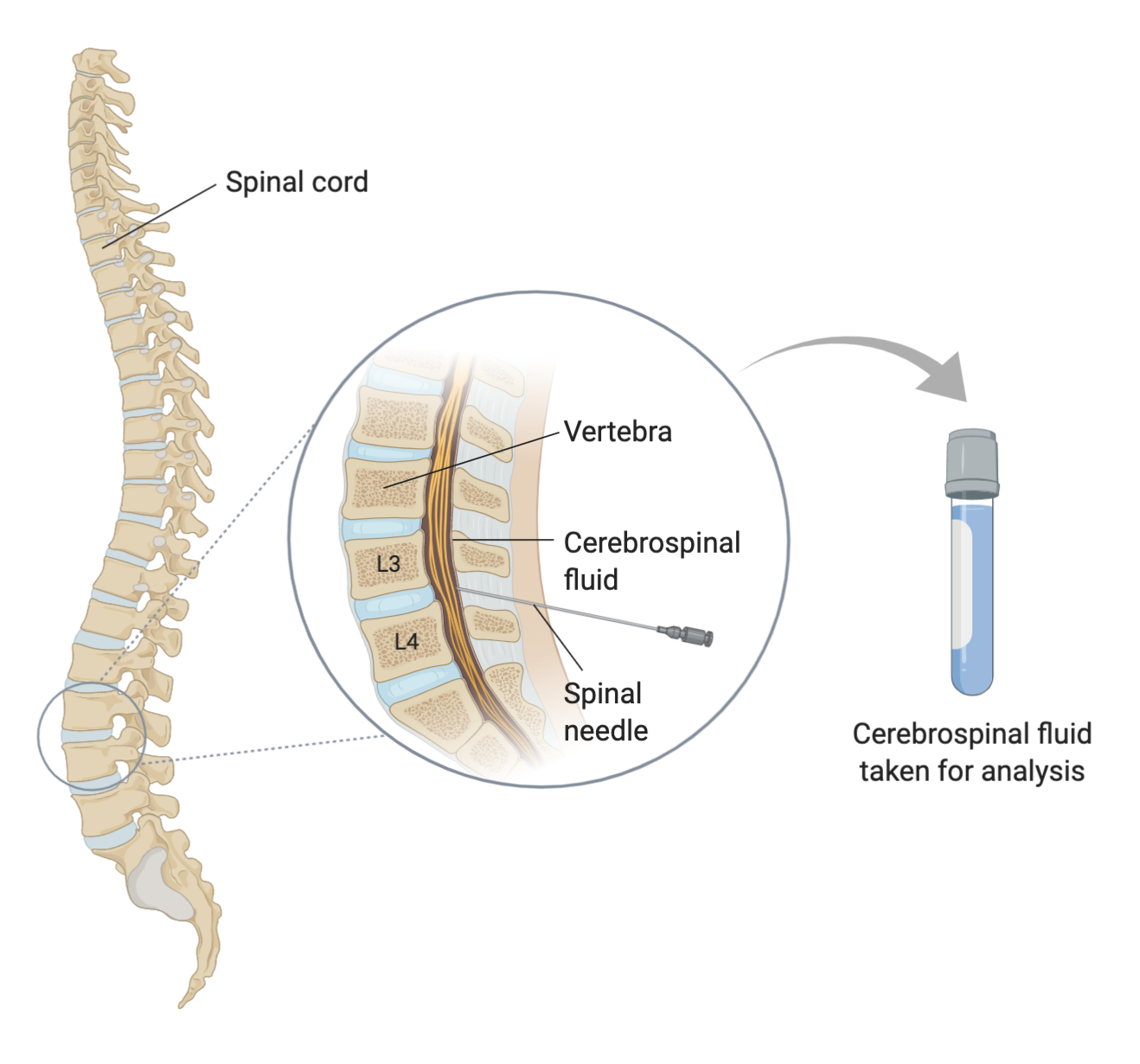
Machine learning models in this field typically undergo several stages of development. First, researchers gather blood samples from patients with confirmed diagnoses through PET scans or cerebrospinal fluid (CSF) analysis. These samples serve as the “ground truth” for training the AI. The model learns to associate specific protein concentrations with each confirmed disease state.
Once trained, the model is tested against “blind” samples—samples where the diagnosis is known but not revealed to the AI. The goal is to achieve high sensitivity (the ability to correctly identify those with the disease) and high specificity (the ability to correctly identify those without it). For a blood test to be clinically viable, it must demonstrate high accuracy across diverse patient populations, accounting for variables like age, sex, and other underlying health conditions.
Comparing current diagnostic methods
To understand the potential impact of AI-driven blood tests, it is necessary to compare them with the existing tools used in neurology today. The following table outlines the primary differences in application, cost, and invasiveness.
| Diagnostic Method | Primary Target | Invasiveness | Relative Cost | Clinical Utility |
|---|---|---|---|---|
| Cerebrospinal Fluid (CSF) Analysis | Amyloid, Tau, Alpha-synuclein | High (Lumbar Puncture) | High | Highly accurate; considered a gold standard for protein detection. |
| PET Imaging | Protein accumulation in brain tissue | Low (Radiotracer injection) | Very High | Excellent visualization of pathology; limited by availability and cost. |
| Cognitive Testing | Symptomatic cognitive decline | None | Low | Identifies impairment but cannot confirm biological cause. |
| AI Blood Test (Experimental) | Proteomic signatures | Low (Standard blood draw) | Low to Moderate | Potential for rapid, scalable, and multi-pathology screening. |
What this means for patient care and precision medicine
The shift toward more precise diagnostics has direct implications for how dementia is managed. Many new pharmaceutical interventions, including monoclonal antibodies designed to clear amyloid from the brain, are highly specific to certain types of pathology. If a patient is misdiagnosed with Alzheimer’s but actually has a different form of dementia, these expensive and potentially side-effect-heavy treatments will not work.
Accurate diagnosis through an AI-enhanced blood test would enable “precision neurology.” This means clinicians can tailor treatments to the specific proteinologies present in an individual’s brain. For patients with mixed dementia, this could mean a combination of therapies designed to address multiple disease pathways simultaneously.
Furthermore, early detection is a critical factor in managing neurodegenerative diseases. Many of these conditions begin changing the brain years before physical symptoms appear. A scalable blood test could facilitate earlier screening in high-risk populations, providing a wider window for interventions that may slow cognitive decline.
Potential barriers to clinical implementation
Despite the promise of this technology, several hurdles remain before it becomes a standard part of clinical practice. One primary concern is the “black box” nature of some machine learning models. For a physician to trust a diagnostic tool, they must understand why the AI reached a specific conclusion. Researchers are currently working on “explainable AI” (XAI) to make these proteomic signatures more interpretable for medical professionals.
Regulatory approval from bodies such as the U.S. Food and Drug Administration (FDA) or the European Medicines Agency (EMA) will require rigorous validation in large-scale, diverse clinical trials. These trials must prove that the blood test remains accurate across different ethnicities, ages, and comorbidities, such as diabetes or cardiovascular disease, which can also alter blood protein levels.
Frequently Asked Questions
Can a blood test replace a brain scan?
Currently, AI-driven blood tests are viewed as screening or supplemental tools rather than total replacements for PET scans or CSF analysis. While they offer a faster and less invasive way to identify potential issues, imaging remains necessary to visualize the exact location and density of protein deposits in the brain.

How accurate are these experimental tests?
Accuracy varies depending on the specific proteins being measured and the machine learning model used. While some single-marker tests (like p-tau217) have shown high accuracy for Alzheimer’s, the multi-protein models designed to detect overlapping dementias are still in the experimental and validation stages.
Who will be able to access these tests?
If these tests receive regulatory approval, they are expected to be accessible through standard clinical laboratories. Because they require only a routine blood draw, they are significantly more scalable than current imaging-based diagnostics, potentially making them available in primary care settings.
The next major checkpoint for this technology will be the publication of large-scale validation studies in peer-reviewed medical journals, which will provide the data necessary for regulatory review and clinical adoption.
How do you feel about the use of AI in medical diagnosis? Share your thoughts in the comments below and share this article with your network.
Related reading