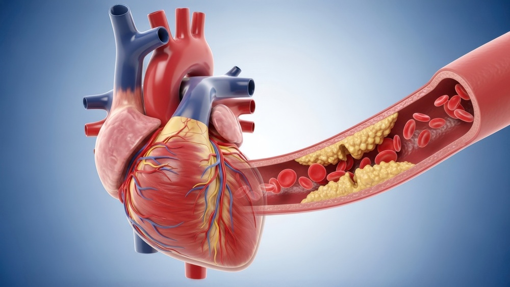
Researchers are increasingly utilizing epicardial fat volume measured via coronary computed tomography angiography (angioTC) as a significant biomarker for identifying subclinical heart failure. By quantifying the adipose tissue surrounding the heart, clinicians may gain earlier insights into cardiac dysfunction before overt clinical symptoms manifest, according to recent findings in cardiovascular imaging research.
As a physician, I often emphasize that the heart’s interaction with surrounding visceral fat is far more dynamic than a simple storage mechanism. Epicardial adipose tissue (EAT) functions as a metabolically active organ, capable of secreting pro-inflammatory cytokines that can directly influence the myocardium. When visualized through angioTC—a standard diagnostic tool for assessing coronary artery disease—this fat layer provides a non-invasive window into the systemic and local metabolic stressors affecting heart function.
The Role of Epicardial Fat in Cardiac Pathology
The relationship between epicardial fat and heart health is rooted in the proximity of these cells to the coronary arteries and the heart muscle itself. According to the American Heart Association, adipose tissue accumulation is frequently associated with metabolic syndrome, which significantly elevates the risk of developing heart failure with preserved ejection fraction (HFpEF). Unlike subcutaneous fat, EAT shares a microcirculation with the underlying myocardium, meaning that inflammatory markers released by this fat deposit can diffuse directly into the heart tissue.
When clinicians perform a coronary angioTC, the primary focus is typically the detection of obstructive plaques. However, the software used for these scans can also quantify the volume and density of the surrounding epicardial fat. A higher volume of EAT is often correlated with increased insulin resistance, hypertension, and systemic inflammation—all of which serve as precursors to structural changes in the heart, such as left ventricular hypertrophy or diastolic dysfunction, as noted in studies published by the European Society of Cardiology.
AngioTC as a Predictive Diagnostic Tool
The utility of angioTC in this context lies in its ability to provide a comprehensive cardiovascular profile in a single session. By analyzing the epicardial fat volume alongside coronary anatomy, doctors can better stratify a patient’s risk profile. While traditional risk factors like cholesterol levels and blood pressure remain essential, the addition of EAT quantification offers a more granular look at the patient’s specific cardiac environment.
According to clinical research, patients who demonstrate higher EAT volumes on angioTC but have no history of heart disease may still be at an elevated risk for developing subclinical heart failure. This “subclinical” stage is critical because it represents a window of opportunity where lifestyle modifications—such as weight management, glucose control, and physical activity—could potentially reverse or slow the progression of cardiac remodeling. The Mayo Clinic highlights that early intervention in patients with asymptomatic structural heart changes is a vital component of modern preventive cardiology.
Clinical Implications and Future Directions
Integrating EAT assessment into routine cardiovascular reporting requires standardized protocols to ensure consistency across different imaging centers. Currently, while the association between EAT and cardiac risk is well-established in clinical literature, the widespread adoption of EAT quantification as a standard clinical metric is still evolving. Cardiologists are evaluating how to best incorporate these measurements into existing diagnostic workflows to ensure that the data leads to actionable patient outcomes.
For patients, this means that future cardiac evaluations may include more detailed assessments of body composition around the heart. By focusing on the metabolic activity of the heart’s environment, the medical community is moving toward a more personalized approach to heart failure prevention. The goal is to shift from reactive treatment—where we intervene after symptoms appear—to proactive management based on the specific anatomical and metabolic markers identified during routine imaging.
Next Steps in Cardiovascular Screening
The next checkpoint for this field involves the validation of standardized software tools that can automatically segment and measure epicardial fat during standard angioTC procedures. As these tools become more accessible, we expect to see more longitudinal data confirming how specific EAT thresholds correlate with the timeline of heart failure development. For those currently undergoing cardiovascular screening, it is recommended to discuss individual risk factors and the relevance of imaging findings with a cardiologist. If you have questions regarding your own cardiovascular health or recent diagnostic results, I encourage you to share your experiences or questions in the comments section below.