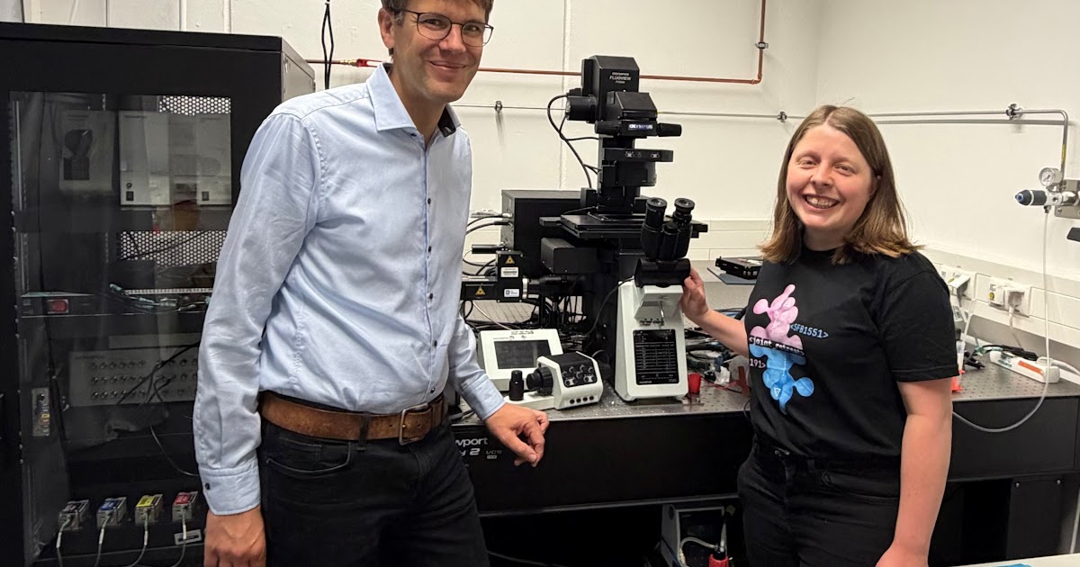
Researchers at Ruhr University Bochum have developed confocal Fast Relaxation Imaging
(cFReI), a new technique to measure protein stability within living cells. By comparing membraneless organelles to the surrounding cytoplasm, the method helps scientists investigate how protein misfolding contributes to neurodegenerative diseases like ALS, Alzheimer’s, and Parkinson’s.
Measuring Protein Stability Inside Living Cells
Scientists have long struggled to observe how proteins behave within the crowded, complex environment of a living cell. To address this, researchers at Ruhr University Bochum, working alongside the Research Center Chemical Sciences and Sustainability, created a method called confocal Fast Relaxation Imaging, or cFReI. This technique allows for the direct measurement of protein unfolding and stability at localized points within a single cell.
The core challenge in understanding neurodegenerative conditions is determining whether specific cellular compartments protect the cell or contribute to disease. Researchers focused their initial analysis on Superoxide Dismutase 1 (SOD1), a protein variant known to accumulate in cellular structures called stress granules. These structures are a type of membraneless organelle (MLO), which are formed through a process known as liquid-liquid phase separation,
a phenomenon compared to the way oil droplets separate in water.
Membraneless Organelles and Neurodegeneration
The role of membraneless organelles in disease remains a subject of intense scientific inquiry. Because these compartments lack a physical membrane, they serve as hubs where proteins and RNA can accumulate. While some organelles are membrane-bound, such as the nucleus and mitochondria, others exist as organelles that are not encased by a membrane,
according to the researchers.
The primary hypothesis being tested is whether these structures act as a shield, sequestering misfolded proteins to protect the rest of the cell, or if they serve as environments that accelerate harmful clumping.
- Amyotrophic lateral sclerosis (ALS)
- Alzheimer’s disease
- Parkinson’s disease
Using cFReI, the team sought to see if the internal environment of these granules altered the stability of the SOD1 protein. The results challenged previous assumptions derived from laboratory-based experiments. Contrary to our expectations from in vitro experiments, we observed that SOD1 is not destabilized within the stress granules,
the researchers noted.
Future Applications for Cellular Research
The ability to map protein stability in real-time within a cell offers a new pathway for drug discovery and medical intervention.
While the initial application utilized SOD1 as a benchmark, the long-term goal is to use cFReI to identify the specific cellular mechanisms that trigger protein misfolding. By pinpointing whether these membraneless organelles are protective or harmful, researchers aim to develop more targeted therapies for neurodegenerative diseases. The research continues to address the significant knowledge gap regarding how these microscopic compartments influence disease progression at the cellular level.
Sources: Nature.
Sources: Sflorg.
Keep reading