The Dawn of Holistic Cellular Imaging: New Microscope Bridges Nano and Micro worlds
Have you ever wondered how scientists observe the intricate dance of life within our cells, tracking everything from massive structural changes to the movements of individual proteins? For decades, researchers have faced a fundamental trade-off: powerful microscopes often excel at visualizing either large cellular components or incredibly small ones, but rarely both concurrently. Now, a groundbreaking innovation from the University of Tokyo promises to shatter that limitation, ushering in a new era of holistic cellular imaging. this isn’t just a technological leap; it’s a potential game-changer for drug discovery, biotechnology, and our fundamental understanding of life itself.
The Challenge of Seeing It All
Since the invention of the microscope in the 16th century, advancements have consistently pushed the boundaries of what we can see. However, these improvements often came with compromises. Two prominent techniques, Quantitative Phase Microscopy (QPM) and Interferometric Scattering (iSCAT) microscopy, exemplify this challenge.
QPM excels at capturing detailed, static images of larger cellular structures – anything over 100 nanometers in size. It achieves this by analyzing how light passes through a sample, revealing subtle differences in density and shape. You can learn more about the principles of QPM at https://www.microscopyu.com/techniques/quantitative-phase-microscopy. However, QPM struggles to detect the smallest particles, like individual proteins.
iSCAT microscopy, conversely, focuses on light that bounces off a sample. This allows it to detect incredibly tiny structures, even single proteins, and track their dynamic movements within living cells. A detailed clarification of iSCAT can be found at https://www.ncbi.nlm.nih.gov/pmc/articles/PMC6288991/.But iSCAT’s focus on back-scattered light limits its ability to provide a broad overview of the cell’s architecture.
Essentially, researchers were forced to choose: a wide-angle view with limited detail, or a close-up on tiny components with a restricted field of vision.
A Revolutionary Solution: Capturing Light From Every Angle
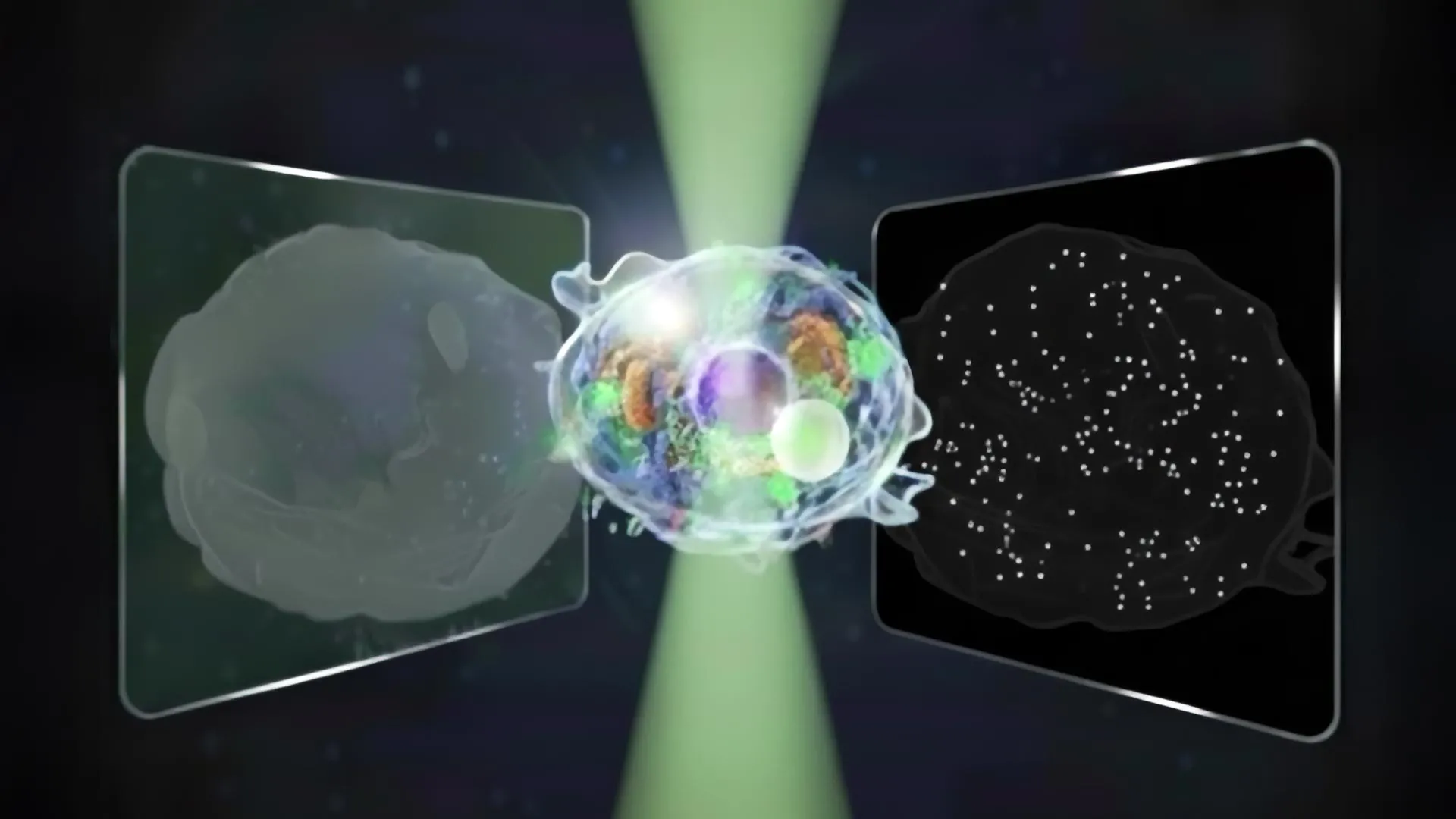
Researchers Kohki Horie, Keiichiro toda, Takuma nakamura, and Takuro Ideguchi at the University of Tokyo have elegantly bypassed this limitation. Their newly developed microscope captures both forward- and back-scattered light simultaneously. This innovative approach,detailed in a recent publication in Nature Communications,allows for the visualization of structures spanning an unprecedented intensity range - fourteen times broader than that of conventional instruments.
“I woudl like to understand dynamic processes inside living cells using non-invasive methods,” explains horie, highlighting the driving force behind the research.
the key to their success lies in a elegant signal separation technique. “Our biggest challenge,” says Toda, “was cleanly separating two kinds of signals from a single image while keeping noise low and avoiding mixing between them.” The team developed a novel algorithm that effectively disentangles the forward- and back-scattered light, providing a comprehensive picture of cellular activity.
Beyond Visualization: Unlocking Cellular Secrets
This isn’t just about seeing more; it’s about understanding more. By analyzing the patterns in both forward- and back-scattered light, the researchers can estimate not only the size of individual particles but also their refractive index – a crucial property that reveals how light interacts with the material, providing insights into its composition and structure.
in a proof-of-concept experiment, the team observed cells undergoing programmed cell death (apoptosis). The microscope successfully captured the movements of both large cellular structures and minuscule particles involved in the death process, offering a holistic view of this complex biological event.
Implications for the Future of Biotechnology and Medicine
The potential applications of this technology are vast. The microscope’s label-free operation – meaning it doesn’t require the use of possibly disruptive dyes – is notably notable. this gentle approach allows for long-term imaging of living cells without causing damage, making it ideal for:
* Pharmaceutical Research: Testing the effects of drugs on cellular processes in real-time, observing subtle changes that might be missed by conventional methods.
* Biotechnology: Monitoring the behavior of cells in bioreactors, optimizing conditions for the production of valuable biomolecules.
* Disease Diagnostics: Identifying and characterizing biomarkers associated with disease, potentially leading to earlier and more accurate diagnoses.
* Exosome and Virus Research: Studying these incredibly small particles –