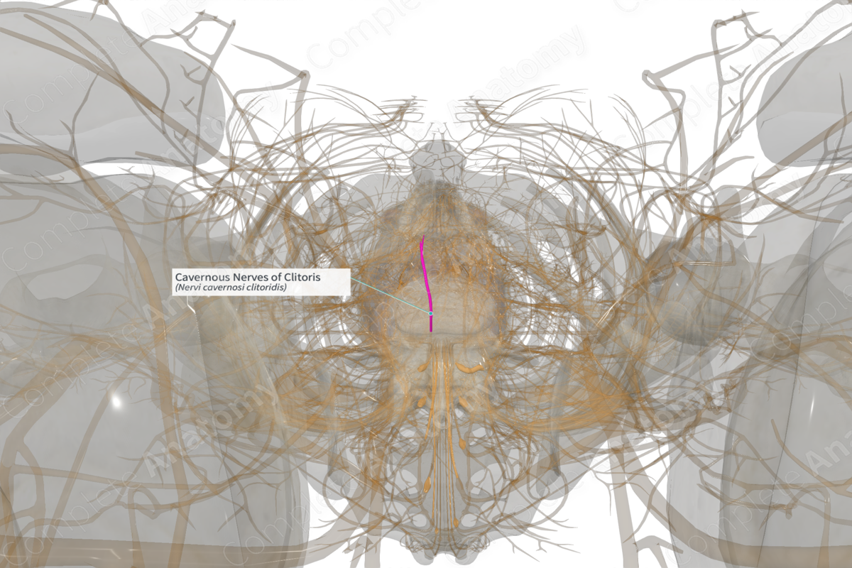
In a landmark advancement for female anatomy, researchers have produced the first-ever 3D map of the clitoris, capturing its complex neural network in high resolution. This breakthrough provides an unprecedented appear at one of the least-studied organs in the human body, offering micron-scale detail that was previously impossible to achieve through traditional medical imaging or dissection.
The study, led by Dr. Ju Young Lee, a postdoctoral researcher at Amsterdam University Medical Centers, utilized a specialized X-ray technique involving a synchrotron—an extremely bright light source—to render the organ’s nerves in three dimensions. This effort was part of the broader Human Organ Atlas initiative, which seeks to map whole human organs in fine detail to improve medical understanding and clinical outcomes via Live Science.
For centuries, the clitoris has been marginalized in scientific literature. Historically described by a 16th-century anatomist as the “shameful member,” the organ has been ignored in research due to social taboos and the physical challenges of dissection, as It’s situated deep within the pelvis and surrounded by other organs. While Magnetic Resonance Imaging (MRI) has previously helped characterize the general structure of the clitoris, those images lacked the resolution necessary to spot the fine nerves responsible for sensitivity.
By imaging two postmortem donated pelvises, the research team was able to visualize the dorsal nerve—the primary sensory nerve of the clitoris—and its intricate branching patterns. This discovery not only fills a critical gap in anatomical knowledge but also highlights a significant historical disparity in medical research, coming nearly three decades after similar mapping was conducted for the penis via HSB Noticias.
High-Resolution Mapping and the Role of Synchrotron Technology
The precision of this new map is the result of synchrotron-based computed tomography. Unlike standard X-rays or MRIs, this technology allows for images at a micrometric scale, enabling scientists to see structures as small as 0.2 to 0.7 mm via El Espectador. This level of detail is essential for understanding how the clitoris functions and how it transmits sensory information.
The researchers discovered that within the glans of the clitoris, the dorsal nerve organizes into several small “trunks” that branch out like a tree, extending toward the surface. This dense, branching architecture explains the organ’s extreme sensitivity. The study further revealed that these nerves do not remain isolated within the clitoris but extend beyond it, providing a more comprehensive view of the pelvic neural network.
Correcting Medical Misconceptions
For years, medical textbooks have provided incomplete or inaccurate information regarding the clitoris. This 3D reconstruction allows the scientific community to identify and correct these errors, fundamentally redefining the existing knowledge of female anatomy. This shift is expected to impact medical education, ensuring that future generations of healthcare professionals are taught using precise, updated data rather than outdated or imprecise diagrams via HSB Noticias.
Clinical Implications for Pelvic Surgery
Beyond the academic value of the map, the findings have immediate practical applications for surgical procedures. A more accurate understanding of the clitoral nerve network is critical for improving the safety and success of pelvic surgeries, such as hysterectomies and reconstructive procedures.
Previously, the risk of nerve damage during these interventions—and the subsequent impact on female sexual function—was often underestimated or poorly addressed in clinical practice. With the new 3D map, surgeons can better identify the exact location of nerves, significantly reducing the risk of accidental damage and helping to preserve sexual function for patients undergoing necessary pelvic operations via HSB Noticias.
Key Takeaways from the Study
- Technological Breakthrough: Used synchrotron X-rays to achieve micron-scale 3D resolution, surpassing the capabilities of MRI.
- Neural Discovery: Identified the dorsal nerve’s branching structure in the glans, with nerve trunks measuring between 0.2 and 0.7 mm.
- Educational Impact: Corrects long-standing inaccuracies in anatomy textbooks and medical training.
- Surgical Benefit: Provides a roadmap for surgeons to avoid nerve damage during pelvic surgeries like hysterectomies.
- Addressing the Gap: Closes a nearly 30-year research gap between the anatomical mapping of the penis and the clitoris.
The project’s integration into the Human Organ Atlas ensures that this data will contribute to a wider global effort to map the human body’s intricacies. As the medical community adopts these findings, the focus shifts toward integrating this high-resolution anatomical data into standard surgical protocols to improve patient outcomes worldwide.
Further updates on the Human Organ Atlas and subsequent applications of this neural mapping in clinical settings are expected as the research is integrated into medical curricula and surgical guidelines.
Do you believe medical education needs a more comprehensive overhaul of anatomical teaching? Share your thoughts in the comments below or share this article with your network.
Keep reading