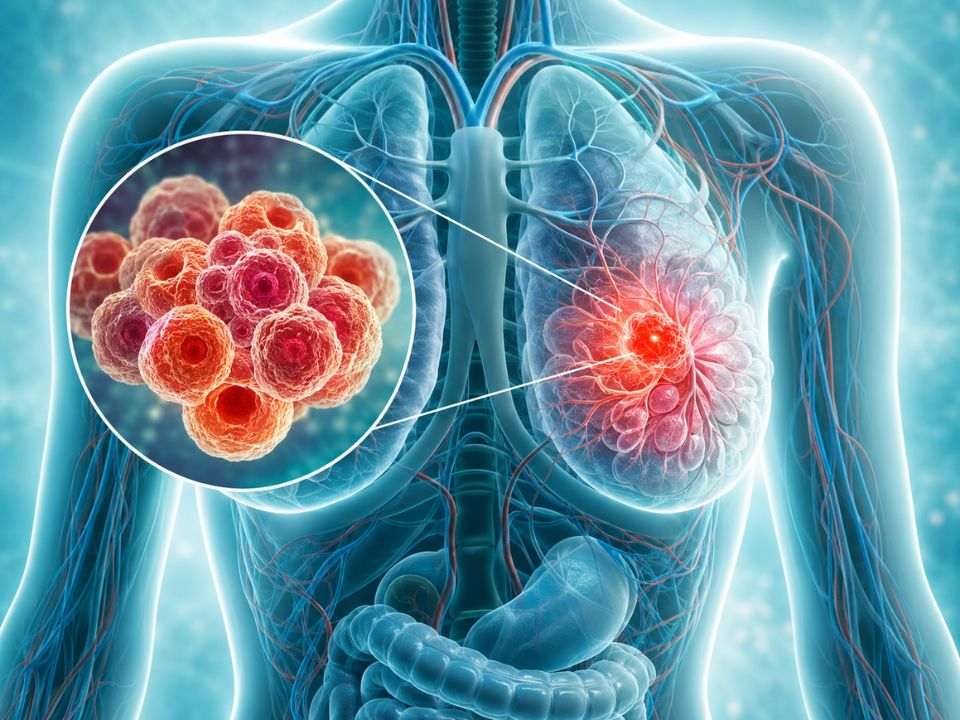
Digital pathology is improving the ability of clinicians to predict patient outcomes in breast cancer by utilizing artificial intelligence to analyze tissue samples with greater precision than traditional microscopy. According to research highlighted by Newslab, these AI-driven tools identify complex morphological patterns and biomarkers that correlate with survival rates and treatment responses, allowing for more personalized therapeutic strategies.
The shift from analog to digital pathology involves scanning glass slides into high-resolution images. Once digitized, machine learning algorithms can quantify cellular structures and spatial arrangements that are often invisible or too subtle for the human eye to measure consistently across thousands of cells. This transition reduces the subjectivity inherent in manual pathology reports and provides a standardized metric for disease progression.
Breast cancer outcomes vary significantly based on the molecular subtype of the tumor. By integrating digital imaging with computational analysis, pathologists can better distinguish between aggressive and indolent tumors. This capability is critical for determining whether a patient requires aggressive chemotherapy or can be managed with less invasive endocrine therapies, directly impacting the quality of life and survival probability for patients.
AI Integration in Breast Cancer Prognosis
Artificial intelligence models in digital pathology operate by breaking down whole-slide images into smaller patches, which the software then analyzes for specific features. According to the National Cancer Institute, breast cancer is not a single disease but a collection of different subtypes, and AI helps map these subtypes by detecting subtle variations in nuclear grade and mitotic figures.
These algorithms can identify “hot spots” of high cellular proliferation, which are often the most aggressive parts of a tumor. While a human pathologist might miss a small area of high-grade cells in a large biopsy sample, AI scans the entire slide, ensuring that the most aggressive elements of the cancer are factored into the prognosis. This comprehensive analysis leads to more accurate staging and a more precise prediction of how the cancer will behave over time.
Beyond simple cell counting, digital pathology allows for the study of the tumor microenvironment. This includes analyzing the presence and location of tumor-infiltrating lymphocytes (TILs). A high concentration of these immune cells often correlates with a better prognosis and a more positive response to immunotherapy, providing a biological marker that guides the selection of specific drugs.
Improving Precision in Biomarker Detection
The detection of biomarkers such as HER2 and ER/PR (estrogen and progesterone receptors) is fundamental to breast cancer treatment. Digital pathology enhances the accuracy of these tests by using automated quantification. According to the American Society of Clinical Oncology, precision in biomarker testing is essential to avoid administering expensive and potentially toxic therapies to patients who will not benefit from them.
Digital tools eliminate the “inter-observer variability” that occurs when two different pathologists interpret the same slide differently. By using a calibrated algorithm, clinics can achieve a level of consistency that ensures a patient receives the same diagnosis regardless of which laboratory processes the sample. This standardization is particularly vital for patients in remote areas who send their slides to centralized reference centers for a second opinion.
Furthermore, the ability to archive these images digitally allows for longitudinal studies. Researchers can revisit the original slides of patients who survived or relapsed over a decade, applying new, more advanced AI models to old data to discover new predictors of recurrence. This creates a continuous feedback loop that improves the predictive power of digital pathology every year.
Clinical Impact and Patient Outcomes
The primary goal of predicting outcomes through digital pathology is the implementation of “de-escalation” of therapy. For patients whose digital signatures indicate a very low risk of recurrence, doctors may be able to safely reduce the dose of chemotherapy or avoid radiation entirely. This reduces the long-term side effects of treatment, such as cardiac toxicity or secondary malignancies.
Conversely, for patients with “hidden” aggressive features detected by AI, clinicians can escalate treatment early. Early intervention with more aggressive protocols for high-risk patients can prevent the cancer from metastasizing, which is the single most important factor in increasing long-term survival rates. The precision offered by digital pathology turns a general diagnosis into a specific roadmap for individual care.
The integration of these tools into standard clinical workflows is currently expanding. While many hospitals still rely on traditional microscopy, the adoption of digital pathology is accelerating as regulatory bodies approve more AI-based diagnostic aids. The transition requires significant investment in high-speed scanners and secure data storage, but the trade-off is a measurable increase in diagnostic accuracy.
The Future of Computational Pathology
The next phase of digital pathology involves “multimodal” integration, where digital slide data is combined with genomic sequencing and radiomics (data from CT or MRI scans). By layering these different data types, clinicians can create a “digital twin” of the patient’s tumor, allowing them to simulate how a specific drug might interact with the cancer before the patient ever receives the first dose.
This approach moves breast cancer care toward a truly predictive model. Instead of treating the cancer based on how it looks today, doctors can predict how it will evolve. The use of deep learning allows these systems to recognize patterns across millions of cases globally, bringing the collective expertise of the world’s best pathologists to every single biopsy slide.
As these technologies mature, the role of the pathologist is shifting from a primary observer to a curator of AI-generated data. The pathologist verifies the AI’s findings and integrates them into the broader clinical context of the patient’s health, ensuring that the human element of medical judgment remains central to the diagnostic process.
The continued rollout of AI-integrated pathology platforms remains the next major checkpoint for healthcare systems globally, with ongoing clinical trials assessing the direct impact of AI-led prognosis on overall survival rates. Patients and providers are encouraged to consult with their oncology teams regarding the availability of digital pathology and biomarker quantification at their specific treatment centers.
Worth a look
- Why Do You Get Sick as Soon as Your Vacation Starts? (Leisure Sickness Explained)
- Ragweed Allergies: How to Identify and Manage This Common Garden Weed
- Wet-Bulb Temperature vs. Heat Index: What Actually Predicts Danger (daybreakwire.com)
- Cataract Surgery Outcomes in Thyroid Eye Disease (archynewsy.com)