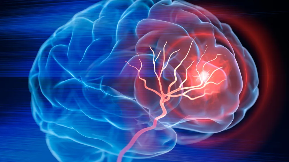
Researchers mapping blood clots in the brain following a stroke have uncovered critical spatial patterns that explain why some patients suffer prolonged cognitive decline while others recover more predictably. According to medical researchers studying cerebrovascular pathology, micro-thrombi and secondary obstructions frequently persist within specific microvascular networks long after acute large-vessel blockages are cleared by mechanical thrombectomy or thrombolysis. This high-resolution mapping effort provides clinicians with a clearer framework for understanding how residual microvascular occlusion impacts post-stroke recovery, shedding new light on persistent neurological deficits.
Strokes remain a leading cause of long-term adult disability worldwide, driven largely by the complex cellular cascades that follow initial ischemia. While acute interventions successfully restore blood flow through major cerebral arteries, recent imaging and histological analyses show that microscopic blood clots often remain trapped in downstream capillary beds. Medical specialists point out that these secondary blockages starve surrounding neural tissue of oxygen and glucose, fueling ongoing inflammation and tissue damage well after the acute emergency has passed.
Understanding the exact topography of these residual blockages changes how researchers approach post-stroke neuroprotection. Rather than focusing solely on the primary site of arterial occlusion, clinical investigators are now evaluating therapies designed to target microvascular health and prevent downstream clotting cascades. This evolving perspective bridges a vital gap in stroke medicine, connecting emergency reperfusion techniques with long-term rehabilitation outcomes.
Mapping Microvascular Obstructions After Acute Stroke
Advanced neuroimaging and post-mortem tissue analysis have allowed medical teams to chart the precise distribution of cerebral blood clots with unprecedented clarity. By examining the micro-architecture of blood vessels following an ischemic event, researchers have identified common accumulation zones where microscopic clots tend to lodge. These locations often correspond to watershed areas of the brain, which are naturally vulnerable to drops in perfusion pressure.
The persistence of these micro-emboli explains a clinical puzzle that has long frustrated physicians: why patients with successful large-vessel recanalization can still experience significant cognitive and motor deficits. When capillary networks remain obstructed, neurons are deprived of essential nutrients despite a clear main artery. Documenting these patterns gives neurologists a tangible anatomical target for designing adjuvant therapies that clear microvascular blockages before permanent scarring occurs.
Clinical Implications for Recovery and Treatment Protocols
Translating these mapping findings into bedside care requires a shift in how hospitals monitor and treat stroke survivors during the critical recovery window. Current standard care prioritizes rapid recanalization of large vessels through medications or catheter-based procedures. However, emerging data suggest that secondary anti-clotting protocols or targeted vasodilators may be necessary to address the microvascular bed comprehensively.
Healthcare providers are also exploring how these persistent clots influence rehabilitation timelines. Patients with extensive microvascular involvement often experience slower cognitive processing speeds and greater fatigue during physical therapy. Recognizing that these symptoms stem from physical capillary blockages rather than generalized weakness helps care teams tailor rehabilitation strategies to the physiological realities of brain tissue recovery.
Next Steps in Cerebrovascular Research
As research groups continue to refine these vascular maps, clinical trials are underway to test drugs capable of dissolving micro-clots without increasing the risk of intracranial hemorrhage. Investigators aim to establish standardized imaging protocols that can identify capillary-level obstructions in living patients, allowing for personalized treatment adjustments during the acute recovery phase. Official updates and clinical trial registries will provide ongoing documentation as these diagnostic tools move toward hospital adoption.
Medical professionals and researchers anticipate that integrating microvascular mapping into routine stroke care will ultimately improve functional outcomes for thousands of patients annually. Readers seeking further details on clinical guidelines and ongoing neurological studies can consult resources provided by the World Health Organization or national neurological research institutes. We welcome your thoughts and insights on these findings in the comments below, and we encourage you to share this article with colleagues and interested readers.
Worth a look
- Early Lung Cancer Screening: How It Works and Why It Matters
- Hair Loss Care Guide: Expert Solutions from Seoul National University Specialists
- University of Copenhagen Study Finds Brain Is Wired to Defend Higher Weight (archyworldys.com)
- 4 Things to Avoid After 6 P.M. for Better Blood Pressure (world-today-news.com)