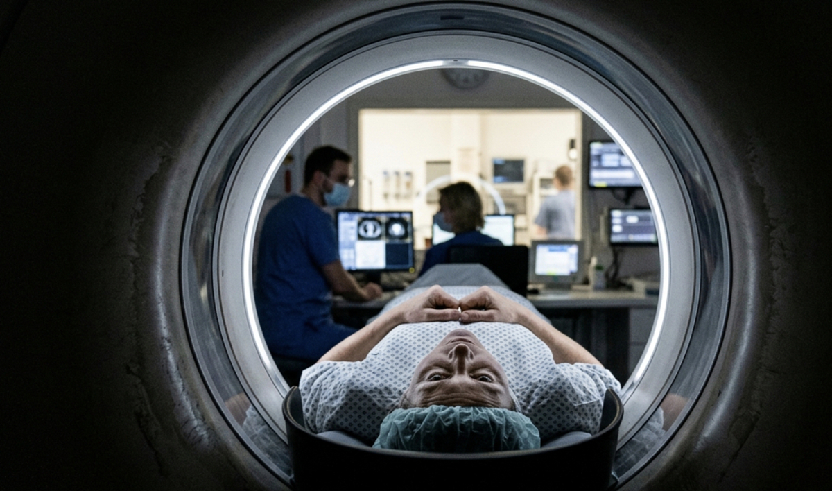
Recent epidemiological research suggests that the long-term cancer risk associated with low-dose ionizing radiation from computed tomography (CT) scans may be higher than previously estimated. While CT scans remain a vital diagnostic tool in modern medicine, providing rapid and precise imaging for trauma, oncology, and internal medicine, the cumulative impact of repeated radiation exposure has become a focal point for public health authorities and radiology experts worldwide.
According to data published by the National Cancer Institute (NCI), CT scans deliver significantly higher doses of radiation than standard X-rays. While a single scan rarely poses a substantial individual risk, the widespread clinical adoption of medical imaging—particularly for asymptomatic screening or repeated monitoring—has prompted researchers to re-evaluate the linear non-threshold (LNT) model, which assumes that any exposure to radiation carries a potential risk of inducing cellular mutations that could lead to malignancy later in life.
The Physics of Risk: Understanding Ionizing Radiation
To understand why researchers are revisiting these risks, one must look at how CT technology functions. Unlike traditional radiography, which captures a single projection, a CT scanner rotates an X-ray source around the body, capturing hundreds of images that are reconstructed into a 3D view. This process inherently requires a higher flux of ionizing radiation to achieve the necessary image resolution.
The International Commission on Radiological Protection (ICRP) emphasizes the principle of “ALARA”—keeping radiation exposure “as low as reasonably achievable.” This framework is designed to balance the immediate diagnostic benefit of a scan against the hypothetical, long-term stochastic effects of radiation, such as DNA damage. When radiation passes through human tissue, it can break chemical bonds in DNA. While cells possess sophisticated repair mechanisms, errors in this process can occasionally result in mutations that accumulate over decades, potentially manifesting as secondary cancers.
Clinical Applications and Patient Safety
In my clinical practice at Charité, we prioritize the necessity of every imaging request. The diagnostic utility of a CT scan in an emergency setting—such as detecting an intracranial hemorrhage or a pulmonary embolism—far outweighs the theoretical lifetime risk of radiation-induced cancer. However, the concern arises in non-urgent scenarios where alternative imaging modalities, such as Magnetic Resonance Imaging (MRI) or ultrasound, could provide the same diagnostic information without the use of ionizing radiation.
The U.S. Food and Drug Administration (FDA) has actively promoted initiatives to reduce patient exposure by encouraging the use of pediatric-specific protocols and dose-modulation technology. These software advancements allow scanners to adjust the radiation output based on the density and size of the patient’s body, effectively lowering the dose while maintaining diagnostic quality. Patients are encouraged to maintain a personal medical imaging record, allowing physicians to track their cumulative lifetime exposure.
Evaluating the Epidemiological Evidence
Recent studies have attempted to quantify the relationship between medical radiation and cancer incidence by linking large-scale hospital databases with national cancer registries. A study published in Nature Medicine analyzed millions of patient records to evaluate the risk of leukemia and other cancers following pediatric CT exposure. The findings indicated a modest but statistically significant increase in risk for those who received high cumulative doses compared to those who did not.
It is important to note that these figures are often expressed as relative risk. For the average patient, the absolute risk of developing cancer from a single CT scan remains extremely low. The discussion is not about abandoning CT technology, but about refining clinical guidelines to ensure that the “benefit-to-risk” ratio remains strictly in favor of the patient’s health. Medical societies, including the American College of Radiology (ACR), continue to update their “Appropriateness Criteria” to help clinicians choose the most effective and safest imaging test for specific patient conditions.
Next Steps for Patient Advocacy
The medical community is moving toward a more transparent dialogue regarding radiation safety. Future developments in artificial intelligence are expected to further reduce scan times and radiation doses by improving image reconstruction algorithms. For patients, the most important step is to engage in shared decision-making with their healthcare providers.
Before consenting to a CT scan, patients should feel empowered to ask their physician: “Is this scan medically necessary for my current condition?” and “Are there alternative imaging options that do not involve ionizing radiation?” Monitoring the upcoming guidelines from the World Health Organization (WHO) regarding medical radiation exposure will provide further clarity as new data becomes available. We invite readers to share their experiences with medical imaging and their thoughts on how healthcare systems can better manage diagnostic radiation in the comments section below.
Worth a look