How Viruses Invade Cells: New Microscopy Reveals a Surprising Cellular ‘Dance’
As winter approaches, the familiar threat of influenza resurfaces. Understanding how influenza viruses infect our cells is crucial for developing effective antiviral strategies. Recent groundbreaking research, utilizing a novel microscopy technique, has revealed a surprisingly active role played by the cells themselves in the viral invasion process – a dynamic interaction far more complex than previously understood.
The Conventional View of Viral Infection
for decades, the prevailing model depicted viral infection as a passive process. Viruses were seen as simply attaching to cells and forcing their way inside. However,this view has been challenged by a collaborative research team from Switzerland and japan,lead by Professor Yohei Yamauchi of ETH Zurich. Their work demonstrates that cells aren’t merely victims; they actively participate in the initial stages of infection, almost ‘helping’ the virus gain entry.
ViViD-AFM: A Revolutionary Imaging Technique
The key to this discovery lies in a new imaging method called virus-view dual confocal and AFM (ViViD-AFM). Previous techniques,like electron microscopy,required fixing cells – essentially killing them – to visualize the intricate details of viral entry. While providing static snapshots, these methods missed the dynamic nature of the process. Fluorescence microscopy offered live imaging, but lacked the necessary resolution to observe the fine-scale interactions.
ViViD-AFM overcomes these limitations by combining the high spatial resolution of atomic force microscopy (AFM) with the live-cell imaging capabilities of fluorescence microscopy. this allows researchers to observe, in real-time and with unprecedented detail, the moment an influenza virus penetrates a living cell.
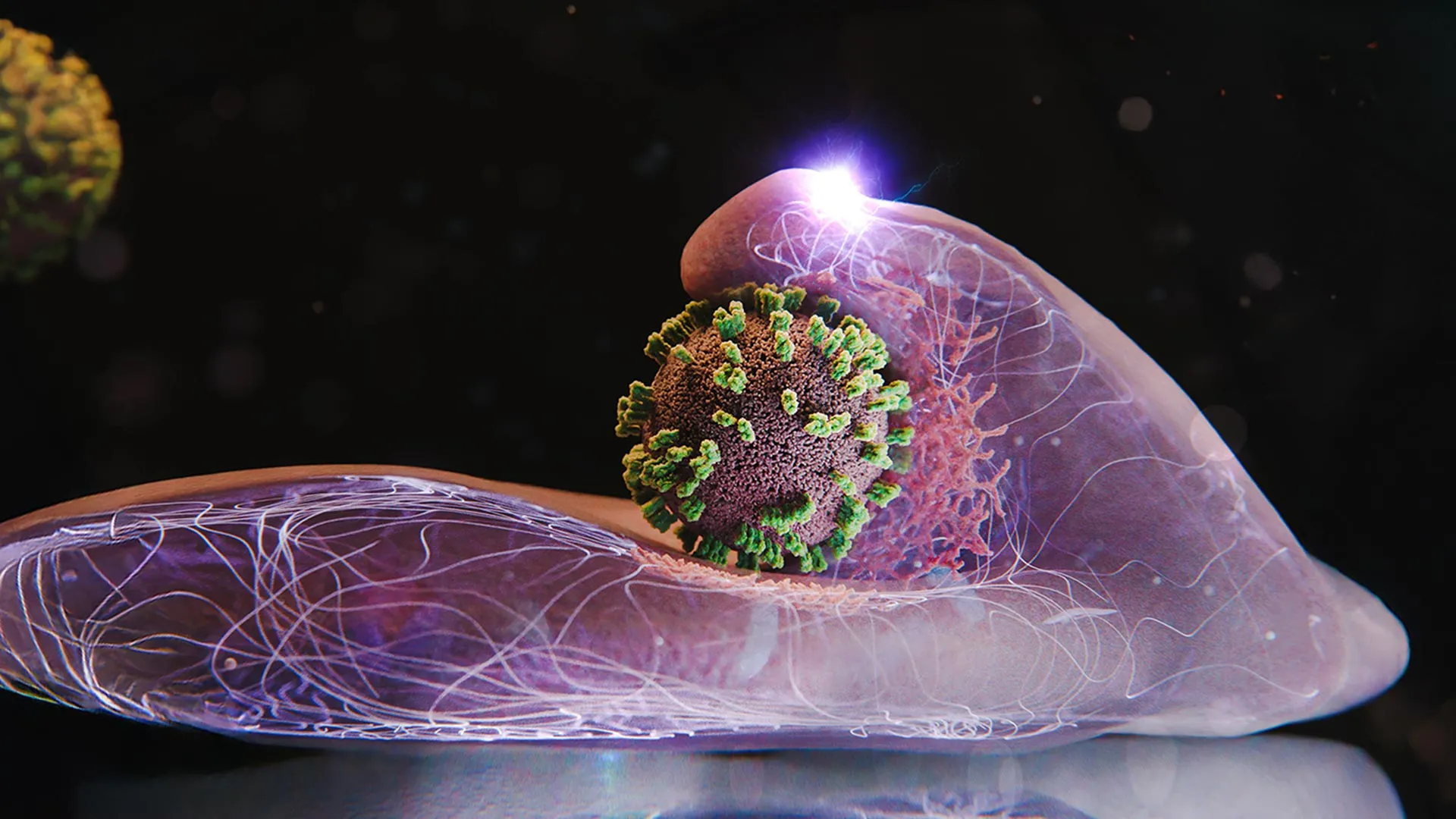
The ‘Dance’ Between virus and Cell: A Step-by-step Process
What the researchers observed was unexpected. Instead of a forceful entry, the process resembles a carefully choreographed ‘dance.’ Here’s a breakdown of the key steps:
- Initial Attachment: The influenza virus initially attaches to specific receptor molecules on the cell surface. This isn’t a random collision; the virus actively ‘surfs’ along the cell membrane, seeking out areas densely populated with these receptors – maximizing its chances of successful entry.
- Membrane invagination: Upon binding to receptors, the cell membrane begins to deform, forming a small indentation. This isn’t a passive response to the virus’s weight; the cell actively initiates this process.
- clathrin’s Role: A structural protein called clathrin plays a critical role in shaping and stabilizing this deepening pocket. The researchers observed the cell actively summoning clathrin proteins to the site of viral attachment, further reinforcing the invagination.
- Vesicle Formation & Internalization: As the pocket expands, it entirely envelops the virus, forming a vesicle – a small bubble-like structure. The cell then pulls this vesicle inward, effectively internalizing the virus. the vesicle’s outer coat dissolves, releasing the virus into the cell’s interior.
- Active Seizure: Remarkably, the researchers observed wave-like motions in the cell membrane, actively pushing upward as if attempting to ‘seize’ the virus and prevent it from drifting away.
Why Cells Facilitate Viral Entry
This seemingly counterintuitive behaviour – a cell actively aiding its own infection – stems from the virus exploiting a pre-existing cellular uptake system. Cells routinely use this system to bring essential substances like hormones, cholesterol, and iron into the cell. The influenza virus cleverly hijacks this system for its own purposes. While the cell doesn’t benefit from being infected, disrupting this uptake system woudl severely compromise its normal function.
Implications for Antiviral Drug Growth & Beyond
The insights gained from ViViD-AFM have critically important implications for antiviral research. The ability to observe viral infection in real-time provides a powerful platform for:
* Direct Drug Testing: Researchers can now directly test the efficacy of antiviral drug candidates in live cell cultures, observing their impact on each stage of the infection process.
* targeted Therapies: Understanding the specific cellular mechanisms involved in viral entry opens the door to developing targeted therapies that disrupt these processes, preventing infection.
* Vaccine Research: The technique can also be applied to study how vaccines interact with cells, providing a real-time view of the immune response.
* Broader Viral Research: ViViD-AFM isn’t limited to influenza; it has the potential to be used to study the entry mechanisms of other viruses, furthering our understanding of viral pathogenesis.
This research represents a paradigm shift in our understanding of viral infection. It highlights the intricate and dynamic interplay between viruses and their host cells, paving the way for more effective antiviral strategies and a deeper understanding of the fundamental processes