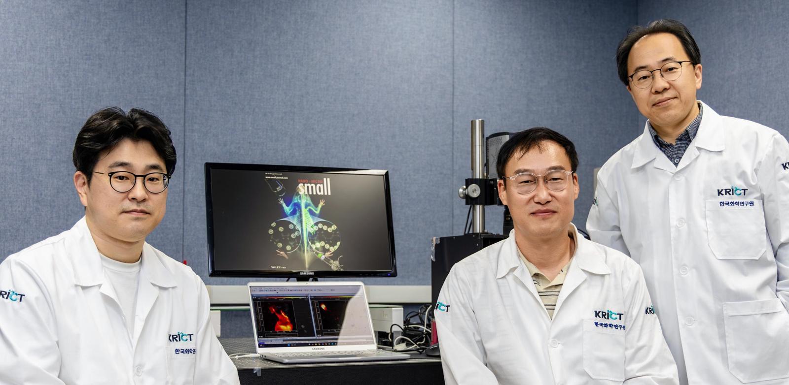
Researchers at the Korea Research Institute of Chemical Technology (KRICT) have announced the development of a next-generation near-infrared (NIR) fluorescent material designed to maintain stable signal intensity during prolonged surgical procedures. This advancement, detailed in recent institutional communications, aims to address the common limitation of signal fading in fluorescence-guided surgery, a technique increasingly used by surgeons to visualize tumors and vital structures in real-time.
As a physician and health journalist, I recognize that the ability to maintain consistent image contrast throughout a multi-hour operation is critical for patient safety. Traditional fluorescent dyes often undergo “photobleaching”—a process where the signal degrades under constant light exposure—which can compromise the accuracy of surgical margins. The development reported by the KRICT team focuses on structural modifications to the dye molecule to enhance its photostability, theoretically allowing for clearer, longer-lasting visualization of deep-tissue targets.
Improving Precision in Fluorescence-Guided Surgery
Fluorescence-guided surgery (FGS) relies on the administration of a contrast agent that accumulates in specific tissues, such as malignant tumors, and emits light when excited by a near-infrared laser. Because NIR light penetrates biological tissue more effectively than visible light, it provides surgeons with a “map” of the surgical field that remains hidden to the naked eye. However, the efficacy of this method is strictly limited by the lifespan of the fluorescent signal.
According to research published in the field of molecular imaging, maintaining a high signal-to-noise ratio is the primary challenge in long-duration oncology surgeries. When a dye loses its fluorescence prematurely, the surgeon may lose the ability to distinguish between healthy tissue and cancerous cells, potentially necessitating a change in approach or increasing the risk of incomplete resection. By engineering a material that resists degradation under continuous illumination, the KRICT team is working to extend the window of clinical utility for these imaging agents.
Technical Advancements in Near-Infrared Fluorescent Dyes
The innovation developed at KRICT involves the synthesis of specialized molecular structures that are less susceptible to the oxidative stress typically caused by high-intensity excitation light. In medical imaging, the shift toward near-infrared wavelengths is driven by the fact that blood and water in the human body absorb less NIR light, reducing background interference and allowing for deeper tissue penetration.
Standard agents currently used in clinical settings are often constrained by their rapid clearance from the body or their vulnerability to chemical breakdown. The research team’s approach emphasizes the development of a molecular framework that remains stable in the complex, aqueous environment of the human body. This stability is essential for procedures that require precise identification of lymph nodes, blood vessels, or tumor boundaries that might be obscured by surrounding anatomical structures.
Clinical Implications for Oncology and Surgical Outcomes
The clinical impact of a more stable fluorescent agent could be significant for oncology departments worldwide. In cases of complex tumor resections, such as those involving the liver or pancreas, the duration of the surgery can exceed several hours. If a contrast agent remains active for the duration of these procedures, surgeons may achieve higher rates of complete tumor excision, potentially reducing the likelihood of local recurrence.
Furthermore, improved imaging stability supports the broader adoption of minimally invasive robotic-assisted surgeries. In these environments, the surgeon relies entirely on the digital feed provided by the imaging system. A consistent, bright signal ensures that the robotic instruments are guided with the highest possible level of anatomical accuracy, minimizing collateral damage to adjacent nerves and vascular systems.
Future Directions and Regulatory Considerations
While the development of a stable NIR fluorescent material is a notable step in medical innovation, the path to clinical integration involves several regulatory and safety milestones. Any new contrast agent must undergo rigorous preclinical testing to evaluate its toxicity profile, metabolic pathway, and clearance rate from the body. Regulatory bodies, such as the Korea Ministry of Food and Drug Safety (MFDS) or the U.S. Food and Drug Administration (FDA), require comprehensive safety data before such materials can be utilized in human trials.
Researchers are currently focusing on ensuring that the material is not only effective but also biocompatible. The next phase of development will likely involve observing how the agent interacts with various tissue types and confirming that it does not induce adverse reactions during long-term exposure. As the field of molecular imaging continues to evolve, the integration of these stable dyes into the surgical suite represents a convergence of chemistry, engineering, and clinical practice aimed at improving patient prognosis.
For those tracking advancements in surgical technology, further updates regarding clinical trial applications and peer-reviewed safety assessments are expected to be published through official institutional channels and medical journals. As we monitor these developments from our research base in Berlin, we will continue to provide analysis on how these innovations translate into standard-of-care improvements for patients globally.
Have you observed advancements in surgical imaging in your clinical practice? Share your insights or questions in the comments section below, and subscribe to our newsletter for the latest updates on medical innovation.
Worth a look