#Particle #physics #heart #medical #advances #cancer
And suffice to say that the tenuous link between the two disciplines did not stop there. Whether it is radiotherapy, magnetic resonance imaging or even radiopharmaceuticals, physics – and particularly particle physics – supports a number of medical innovations, some of which arise directly from close collaboration between hospitals, doctors and physicists at CERN in Geneva.
Better quality images of the human body
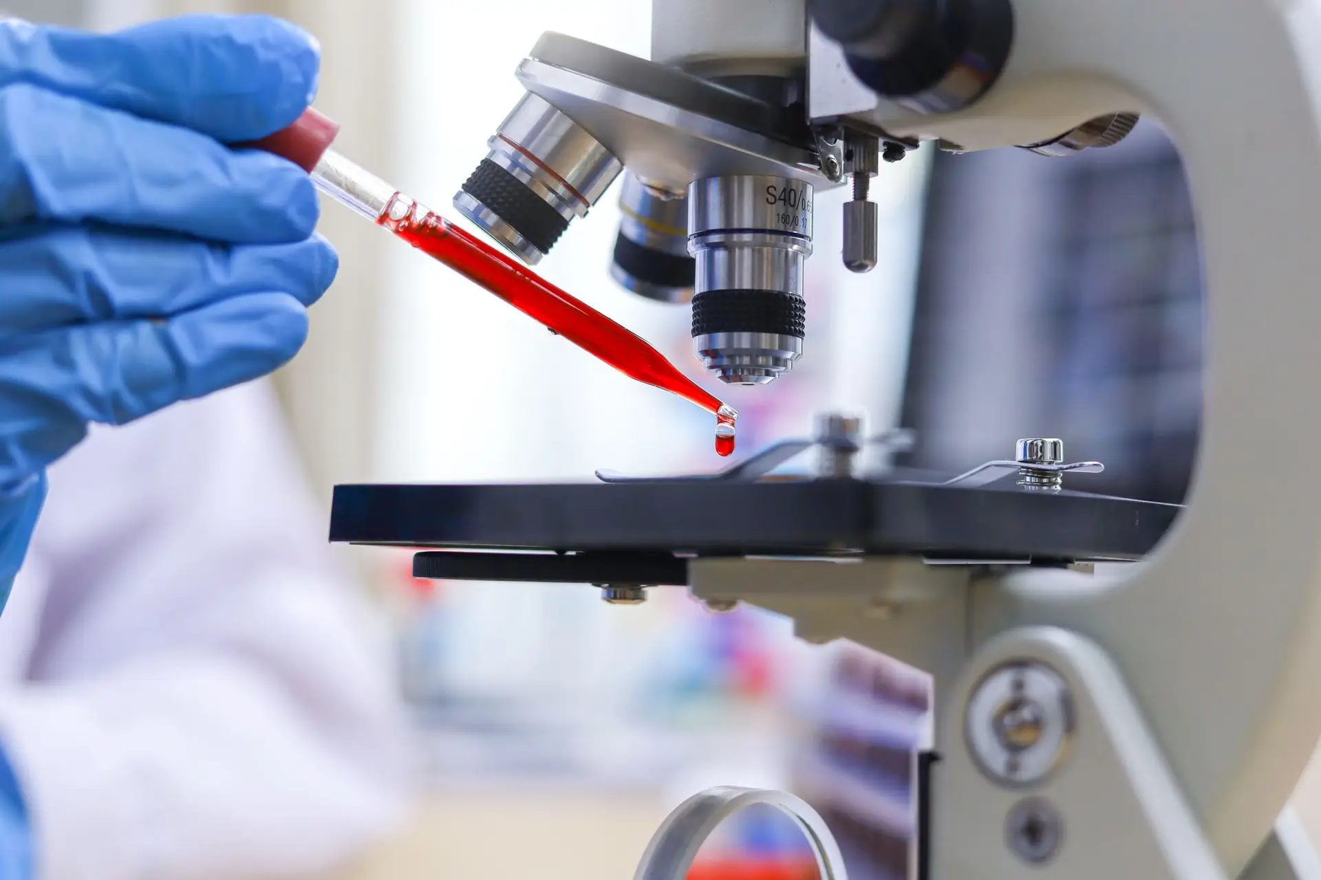
To say more about this collaboration, the case of oncology is undoubtedly one of the most striking examples. Starting with the improvement of what we call positron emission imaging (PET or PET-scan in English), which has now become a common clinical tool for detecting tumors and metastases.
Nearly 1,300 different isotopes of 73 chemical elements are produced at CERN. For their part, the HUG has its own cyclotron, a small accelerator used to produce radioisotopes for diagnostic purposes. “The idea is to use a molecule, such as glucose, and to mark it with radioisotopes by producing a radiopharmaceutical which will be injected into the patient,” explains Professor Valentina Garibotto, head doctor of the medicine department. nuclear and molecular imaging of HUG. One of the fascinating and unique characteristics of this technique is that we can see all the interactions between the radiopharmaceutical and the body molecules it targets, without causing damage to healthy tissues.
Flashback to the 1980s. Professor Antoine Geissbuhler, dean of the Faculty of Medicine at the University of Geneva and director of teaching and research at the University Hospitals of Geneva (HUG), was still a medical student when he landed a job in the nuclear medicine department of HUG. Anyone who is already passionate about computer programming is far from imagining that this commitment will allow him to collaborate with two renowned physicists: Georges Charpak, recipient of the Nobel Prize in 1992 for his invention of the multi-wire proportional chamber – a detector of a new kind capable of recording millions of particle trajectories per second –, and David Townsend, recognized for his innovative work on PET imaging and, later, for his invention of PET-CT, a combination of a PET camera and an X-ray scanner.
Also read: CERN comes out of its bubble
“Physicists from CERN – including David Townsend – had the idea of using the technology developed by Georges Charpak to detect electromagnetic radiation in the context of PET scanning,” describes the Geneva doctor. Result: higher quality images while reducing the necessary radiation dose. And a technology that is very quickly making its inroads into hospitals, particularly at HUG. “The collaboration between CERN and the University Hospitals of Geneva lasted around ten years in order to continue to develop these tools,” recalls Antoine Geissbuhler. It is an example of successful technology transfer to medicine and has become a success story major player in the world of medical imaging.”
Un diagnostic hyperprécis
Let’s stay on the side of particle detectors, but this time to address recent advances in the field of nuclear medicine. The objective: to use small quantities of radioisotopes in order to diagnose and treat cancers. Radioisotopes are isotopes of chemical elements that exhibit nuclear instability, i.e. they spontaneously decay by emitting radioactivity. Some have a lifespan of a few fractions of a second, others of a few billion years.
Also read: Blood tests to detect cancer: where are we?
Why is this technique interesting in oncology? “Tumor sites, like infections, have increased glucose consumption,” replies the doctor. We can thus see very small lesions and diagnose tumors very early that we would not see with other techniques.” Today, dozens of diagnostic radiopharmaceuticals are commonly used in the clinic, particularly for prostate, thyroid and breast cancer.
Bombarding tumors
Furthermore, radiopharmaceuticals are also studied for their therapeutic properties. “Certain isotopes produce high energies and have the characteristic of destroying the cancerous tissues in which they accumulate,” notes Valentina Garibotto. It’s as if we were locally bombarding the tumor pathology. This approach has been around for a long time for the treatment of thyroid cancer. It has also shown greater effectiveness than any other approach to cancer treatment.”
Other therapeutic radiopharmaceuticals have also proven themselves in recent years, such as those targeting a molecule called PSMA (Prostate Specific Membrane Antigen). These use the same principle for metastatic prostate cancer, when the patient does not respond to standard lines of treatment. “Publications have shown the advantage of this approach compared to hormonal therapy or chemotherapy. Large-scale studies are underway and encouraging for use in earlier phases of the disease,” concludes Valentina Garibotto.
Read more: Prostate cancers will increase significantly
Since 2017, the CERN project called Medicis (for Medical Isotopes Collected from Isolde) has brought together an international collaboration including French-speaking hospitals and other European institutes, and aims to develop radioisotopes with an ideal emission profile for use medical. “We have been producing isotopes for nuclear research for more than fifty years and it happens that some of them present very interesting characteristics for medicine, in particular for new targeted therapies,” recalls Manuela Cirilli, head of medical applications at CERN in the technology transfer group. The industry cannot afford to carry out this type of approach without having already clearly identified commercial prospects, as this could prove very costly, which is why CERN acts as a bridge. We are not aiming to achieve industrial production of these isotopes, but can provide the technology to institutions capable of taking over to produce them on the necessary scale.”
Short but powerful radiations
Let’s now move on to particle accelerators. The mention of this term immediately brings to mind the Large Hadron Collider (LHC) with its 27 kilometers in circumference. However, there are also smaller accelerators, which could be modified to deliver radiotherapy in hospitals.
Also read: The giant CERN accelerator project is reinforced
The principle – destroying cancer cells by exposing them to high doses of radiation – has been known for a long time. But it could well prove to be even more efficient in the future. Professor Marie-Catherine Vozenin, head of the Radio-oncology and Radiobiology sector at HUG, is one of the pioneers – with Vincent Favaudon of the Institut Curie in Paris – of what is called Flash radiotherapy. The principle of this concept, discovered around ten years ago, is to irradiate tumors in an ultra-short time, which reduces damage to nearby tissues.
“If we could increase the doses delivered in standard radiotherapy, it would be possible to eradicate all tumors,” describes Marie-Catherine Vozenin. Unfortunately, this has the corollary of inducing toxicity on healthy tissues. This is currently the main limitation of radiotherapy. With Flash, we were able to show, during studies on mice, that it was possible to reduce these complications by using beams capable of delivering doses in a time frame of the order of milliseconds, compared to several minutes with the conventional method.”
In collaboration with CERN – and its particle accelerator called Clear – the researcher and her team have managed to further increase speed and efficiency, with doses of irradiation administered in a few nanoseconds (a billionth of a second), and even in one picosecond (one trillionth of a second). “Healthy tissues are able to manage this very rapid radiation, but not the tumors which are destroyed,” adds Marie-Catherine Vozenin.
Feasibility studies are currently being carried out on patient animals, in particular on cats treated at the Zurich veterinary school for tumors with a poor prognosis. “The experiments show very good anti-tumor effectiveness for all types of tumors, and protection for all types of healthy tissues,” observes the researcher. However, we also see, in certain cases, late complications. This tells us the limits to be defined in order to be able to move forward and conduct clinical trials in humans in the future.”
Although it will probably still take a few years before this technique enters the clinic, this method is already being emulated, since all major cancer centers are developing Flash therapy projects, from the United States to Europe, including China. or Japan.
Finally read: Anticipating the quantum computer revolution: an institute is tackling it at CERN


:quality(85)/cloudfront-us-east-1.images.arcpublishing.com/infobae/UDFZUNW2WNBXMZYMX7RENN5ST4.jpg)