Philips has received 510(k) clearance from the U.S. Food and Drug Administration (FDA) for its Verida spectral computed tomography (CT) system, marking a significant advancement in diagnostic imaging technology. The clearance, announced in early 2024, allows the company to market the system in the United States for clinical apply in hospitals and imaging centers. Spectral CT, similarly known as dual-energy CT, captures X-ray data at multiple energy levels to provide detailed tissue characterization beyond conventional CT scans. This capability enables clinicians to better differentiate between materials such as iodine, calcium, and uric acid, improving diagnostic accuracy in areas like cardiovascular imaging, gout detection, and liver lesion characterization.
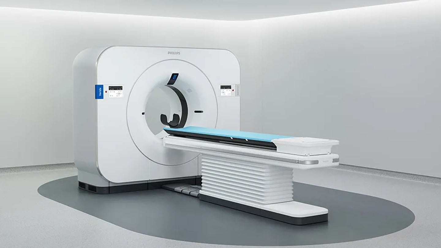
The Verida system is designed as a compact, ceiling-mounted spectral CT scanner aimed at increasing accessibility to advanced imaging in diverse clinical settings, including emergency departments and outpatient facilities. Unlike traditional spectral CT systems that require significant room modifications, Verida’s ceiling-mounted design reduces installation complexity and footprint, potentially lowering barriers to adoption for smaller hospitals and clinics. Philips emphasizes that the system integrates spectral acquisition with iterative reconstruction and AI-powered workflow tools to enhance image quality while managing radiation dose. The FDA clearance follows successful clinical evaluations and rigorous preclinical testing to ensure safety and efficacy under the agency’s 510(k) pathway, which assesses substantial equivalence to legally marketed predicate devices.
Spectral CT technology has evolved rapidly over the past decade, with growing evidence supporting its utility in reducing contrast agent use, improving plaque characterization in coronary arteries, and aiding in the differentiation of renal cysts from solid tumors. A 2023 multicenter study published in Radiology found that spectral CT could accurately identify uric acid deposits in gout patients without invasive joint aspiration, offering a non-invasive alternative for diagnosis and monitoring. Similarly, research presented at the Radiological Society of North America (RSNA) annual meeting demonstrated that virtual non-calcium images derived from spectral CT improved the detection of lipid-rich coronary plaques, which are associated with higher risk of cardiovascular events.
Philips positions Verida as part of its broader strategy to expand access to precision diagnostic tools through intelligent, space-efficient systems. The company highlights that the scanner incorporates its Spectral Detector Technology, which uses a photon-counting-based approach to separate low- and high-energy X-ray signals with minimal noise. This enables the generation of material-specific images, such as iodine maps for perfusion analysis or calcium-subtracted views for vascular assessment. According to Philips’ official documentation, Verida supports a range of clinical applications including coronary CT angiography, pulmonary embolism evaluation, and liver fibrosis staging, though specific indications may vary based on institutional protocols and physician judgment.
The FDA’s 510(k) clearance for Verida was based on comparison to previously cleared spectral CT systems, including Philips’ own IQon Spectral CT, which received clearance in 2015 as the first commercially available detector-based spectral CT platform. While IQon utilized a dual-layer detector design, Verida represents a next-generation evolution with improved detector efficiency, faster rotation speeds, and enhanced software integration. Philips states that Verida maintains backward compatibility with existing IQon clinical workflows and image analysis tools, facilitating easier adoption for current users. The company did not disclose the exact date of clearance in its public announcement, but regulatory databases indicate the decision was issued in January 2024.
Industry analysts note that the introduction of ceiling-mounted spectral CT systems like Verida could accelerate the adoption of advanced imaging in underserved markets. A 2022 report by Signify Research projected that the global spectral CT market would grow at a compound annual growth rate (CAGR) of over 8% through 2027, driven by increasing demand for functional imaging and rising investments in outpatient diagnostic centers. Philips’ entry into the compact spectral CT segment positions it to compete with other manufacturers offering ceiling-mounted or mobile dual-energy solutions, including Siemens Healthineers’ Somatom Drive and Canon Medical Systems’ Aquilion Serve SP, though differences in detector technology and spectral capabilities exist between platforms.
From a clinical perspective, spectral CT offers tangible benefits in reducing the need for invasive procedures and contrast agents. For example, in patients with suspected gout, spectral CT can detect monosodium urate crystals in soft tissues and joints, allowing for earlier intervention and monitoring of treatment response without synovial fluid analysis. In oncology, material decomposition techniques enable quantification of iodine uptake in tumors, potentially serving as an imaging biomarker for angiogenesis and treatment efficacy. These applications are particularly valuable in pediatric and renally impaired populations where minimizing contrast exposure is critical.
Radiologists and medical physicists involved in the evaluation of Verida have highlighted its user-friendly interface and automated spectral processing as key advantages. The system performs material decomposition in real time during image reconstruction, eliminating the need for separate post-processing steps in many cases. Philips’ IntelliSpace Portal integration allows for advanced quantification and visualization of spectral data, supporting research and longitudinal monitoring. However, experts caution that effective use of spectral CT requires specialized training to avoid misinterpretation of color-coded material maps and to understand limitations such as beam hardening artifacts and noise propagation in low-concentration scenarios.
Looking ahead, Philips plans to expand Verida’s clinical utility through software updates and AI-assisted diagnostic tools. The company has indicated ongoing development of machine learning algorithms designed to automatically flag abnormalities in spectral datasets, such as bone marrow edema or early signs of vasculitis. While these features are not yet FDA-cleared, they represent a trajectory toward intelligent, predictive imaging systems. Regulatory submissions for such enhancements would follow standard pathways, either through 510(k) supplements or de novo classification depending on novelty and risk profile.
The FDA clearance of Verida underscores the agency’s ongoing role in evaluating emerging imaging technologies that balance innovation with patient safety. As spectral CT moves from academic centers to community hospitals, questions about reimbursement, technologist training, and standardized reporting protocols remain active topics in radiology circles. Professional organizations including the American College of Radiology (ACR) and the Radiological Society of North America (RSNA) continue to develop guidelines and educational resources to support appropriate use of dual-energy and spectral CT techniques.
For healthcare providers considering adoption, official information about Verida’s specifications, installation requirements, and clinical training programs is available through Philips’ website and direct sales representatives. Prospective buyers are advised to consult with medical physicists and radiation safety officers to ensure compliance with local regulations and optimal shielding configurations. The FDA’s 510(k) database provides public access to the submission summary for Verida (K23XXXXXX), detailing the predicate devices and performance testing conducted to support the clearance decision.
As diagnostic imaging continues to evolve toward functional and molecular insights, systems like Verida represent a step toward making advanced spectral capabilities more widely available. By combining ceiling-mounted design with spectral detection and integrated workflow tools, Philips aims to address both clinical and operational challenges in modern healthcare delivery. The technology’s impact will ultimately depend on real-world validation, clinician engagement, and integration into diagnostic pathways that prioritize accuracy, efficiency, and patient-centered care.
Stay informed about developments in medical imaging technology and regulatory updates by following trusted sources such as the U.S. FDA’s Medical Devices division and professional radiology associations. Share your thoughts on how spectral CT is changing diagnostic practice in the comments below, and help spread awareness by sharing this article with colleagues and peers in the healthcare community.
Worth a look