Revolutionary Imaging Technique Combines Ultrasound and Light for Detailed 3D Views of the Human Body
Researchers from Caltech and the University of Southern California (USC) have unveiled a groundbreaking medical imaging technique poised to transform diagnostics and treatment monitoring. This innovative approach generates rapid, three-dimensional, full-colour images that together reveal both the physical structure of soft tissues and the dynamic function of blood vessels within. Already successfully tested on human subjects, the technology holds immense promise for advancements in areas like breast cancer imaging, diabetic neuropathy assessment, and neurological research.
The findings, recently published in Nature Biomedical Engineering, address critical limitations inherent in currently available imaging modalities. While standard ultrasound is widely accessible and cost-effective, it primarily provides two-dimensional images with a restricted field of view. Photoacoustic imaging, which utilizes laser light to generate sound waves revealing blood vessel activity, excels at visualizing blood flow but struggles to capture complete tissue structure. Established techniques like computed tomography (CT) and magnetic resonance imaging (MRI) often involve trade-offs, including the need for contrast agents, exposure to ionizing radiation, high costs, and lengthy examination times.
Bridging the Gap with RUS-PAT
To overcome these challenges, the research team developed RUS-PAT - an acronym for rotational ultrasound tomography (RUST) combined with photoacoustic tomography (PAT).PAT, originally pioneered over two decades ago by Lihong Wang, Bren Professor of medical Engineering and Electrical Engineering and the Andrew and Peggy Cherng Medical engineering Leadership Chair at Caltech, relies on the principle that tissue molecules vibrate and emit acoustic signals when struck by short laser pulses. These signals are then converted into detailed images.
“The goal was to merge the strengths of ultrasound and photoacoustic imaging,” explains Wang,who also serves as Caltech’s executive officer for medical engineering. “But it’s not like one plus one. We needed to find an optimal way of combining the two technologies.”
The key to this synergy lies in a simplified and more practical design. Customary ultrasound systems require numerous transducers for sound wave transmission and reception, complicating integration with photoacoustic imaging and increasing costs. Recognizing that photoacoustic imaging only requires ultrasound detection, Wang’s team conceived a novel approach: mimicking the light excitation of ultrasound waves in photoacoustic tomography, but utilizing ultrasound itself.
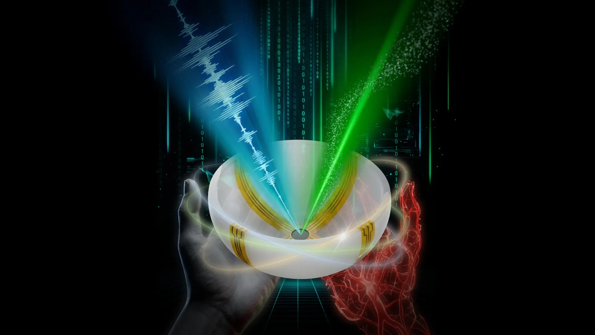
Instead of lasers, a single, wide-field ultrasound transducer generates sound waves throughout the tissue. The same detectors then capture signals from both imaging methods. This streamlined system employs a small number of arc-shaped detectors rotating around a central point, effectively functioning as a full hemispheric detector while considerably reducing complexity and expense.
Demonstrated Clinical Potential
“the novel combination of acoustic and photoacoustic techniques addresses many of the key limitations of widely used medical-imaging techniques in current clinical practice, and, importantly, the feasibility for human request has been demonstrated here in multiple contexts,” states Dr. Charles Y. Liu, a co-author of the study, visiting associate in biology and biological engineering at Caltech, professor at the Keck School of Medicine of USC, director of USC’s Neurorestoration Center, and chair of neurosurgery at the rancho los Amigos National rehabilitation Center.
The versatility of RUS-PAT, enabled by its ability to function wherever light can reach, opens doors to a wide range of clinical applications. In breast cancer imaging, it could facilitate precise tumor localization alongside insights into its biological activity. For patients suffering from diabetic neuropathy, the technique promises simultaneous monitoring of nerve structure and oxygen supply. Furthermore, the technology holds significant potential for brain research, allowing scientists to observe brain anatomy and blood flow dynamics concurrently.
Currently, the system can image tissues up to approximately 4 centimeters deep. The use of endoscopic tools to deliver light could extend this reach to deeper anatomical regions.Importantly, each RUS-PAT scan can be completed in under one minute.
the system, currently configured with ultrasound transducers and a laser positioned beneath a scanning bed, has undergone successful testing on both human volunteers and patients and is now progressing towards broader clinical implementation.
Study Details and Funding
The research was led by Yang Zhang,Shuai na,and Dr. Jonathan J. Russin. Additional contributors from Caltech included Karteekeya Sastry, Li Lin, Junfu Zheng, Yilin Luo, Xin Tong, Yujin An, Peng Hu, and Konstantin Maslov. Dr. Tze-Woei Tan from the Keck School of Medicine of USC also contributed to the study. The research was supported by funding from the National Institutes of Health.
Worth a look