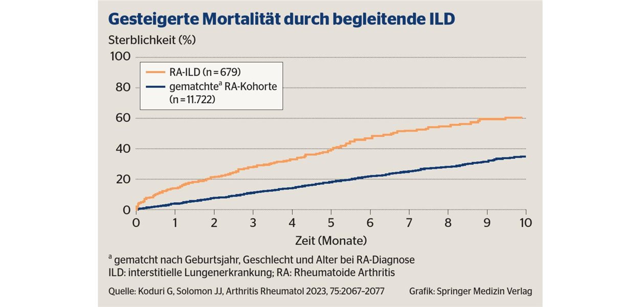
Rheumatoid arthritis (RA) is a systemic autoimmune disease that can cause interstitial lung disease (ILD), a condition where inflammation and scarring affect the lung tissue. According to the American Lung Association, RA-associated ILD can lead to significant respiratory impairment and is a leading cause of morbidity and mortality in patients with the joint disease.
While RA is primarily recognized for attacking the synovial joints, the inflammatory process often extends beyond the musculoskeletal system. Medical research indicates that lung involvement can occur even in the absence of respiratory symptoms, making proactive screening essential for long-term patient survival. Early detection of pulmonary manifestations allows clinicians to adjust immunosuppressive therapies and prevent irreversible fibrosis.
The risk of developing lung complications is higher in patients with specific biomarkers, such as anti-citrullinated protein antibodies (ACPA) and rheumatoid factor (RF). These markers often correlate with a more aggressive disease course and a higher likelihood of extra-articular manifestations, including the lungs, heart, and eyes.
Identifying Rheumatoid Arthritis-Associated Interstitial Lung Disease
Interstitial lung disease in RA patients typically manifests as inflammation or fibrosis within the alveoli and the surrounding tissue. According to the Mayo Clinic, the most common pattern seen in RA is Usual Interstitial Pneumonia (UIP), which is characterized by a “honeycombing” appearance on high-resolution computed tomography (HRCT) scans. Other patterns include Non-Specific Interstitial Pneumonia (NSIP), which often responds more favorably to treatment.
Patients may initially present with subtle symptoms, such as a persistent dry cough or shortness of breath during physical exertion (dyspnea). Because these symptoms often overlap with general aging or cardiovascular issues, they are frequently overlooked during routine rheumatology visits. Clinical guidelines suggest that any RA patient reporting new respiratory symptoms should undergo immediate pulmonary function tests (PFTs) and imaging.
The role of HRCT is critical because standard chest X-rays often fail to detect early-stage ILD. High-resolution imaging allows radiologists to identify early reticulation and ground-glass opacities, which are indicators of active inflammation that can potentially be reversed with aggressive medication.
The Impact of Smoking and Environmental Triggers
Tobacco use significantly exacerbates the risk of lung damage in patients with rheumatoid arthritis. Data from the Johns Hopkins Arthritis Center indicates that smoking not only increases the likelihood of developing RA but also accelerates the progression of ILD. The combination of systemic autoimmunity and inhaled toxins creates a synergistic effect that damages the delicate lung parenchyma.
Environmental exposures, including silica dust and other industrial pollutants, also play a role. Patients working in construction, mining, or manufacturing may face a higher risk of developing “Caplan’s syndrome,” a rare condition characterized by the presence of rheumatoid nodules in the lungs alongside pneumoconiosis.
Cessation of smoking is considered one of the most effective non-pharmacological interventions to slow the progression of lung scarring. When combined with disease-modifying antirheumatic drugs (DMARDs), smoking cessation improves the overall prognosis and reduces the frequency of acute exacerbations.
Treatment Strategies and Medication Risks
Managing RA-associated ILD requires a multidisciplinary approach involving rheumatologists and pulmonologists. The primary goal is to suppress the overactive immune response that targets the lungs. According to the American College of Rheumatology, the use of corticosteroids and immunosuppressants like mycophenolate mofetil or azathioprine is often necessary to stabilize lung function.
However, some medications used to treat joint inflammation can have complex effects on the lungs. Methotrexate, a gold-standard DMARD for joint disease, has been linked in rare cases to methotrexate-induced pneumonitis. This hypersensitivity reaction is distinct from RA-ILD and requires immediate discontinuation of the drug and administration of high-dose steroids.
Recent clinical focus has shifted toward the use of antifibrotic agents, such as ninted danib, which were originally developed for idiopathic pulmonary fibrosis. Research published in the New England Journal of Medicine suggests that antifibrotics can slow the decline of forced vital capacity (FVC) in patients with progressive fibrosing ILD, regardless of the underlying cause, including RA.
Monitoring and Long-term Prognosis
Regular monitoring is the only way to track the progression of lung disease in RA patients. Pulmonologists typically utilize spirometry to measure the volume of air a patient can exhale. A significant drop in the FVC or a decrease in the diffusing capacity for carbon monoxide (DLCO) often signals a need for a change in therapeutic strategy.
The prognosis for RA-ILD varies widely based on the histological pattern. Patients with the NSIP pattern generally have a better prognosis and a more positive response to steroids. In contrast, those with the UIP pattern face a higher risk of progressive fibrosis and respiratory failure, mirroring the trajectory of idiopathic pulmonary fibrosis.
Early intervention remains the most critical factor in improving outcomes. When lung involvement is detected in the “inflammatory phase” before it transitions to the “fibrotic phase,” the potential for preserving lung function is significantly higher.
Frequently Asked Questions
Can rheumatoid arthritis cause permanent lung scarring?
Yes. If the inflammation in the lungs is not controlled, it can lead to fibrosis, which is the formation of permanent scar tissue that makes it harder for the lungs to exchange oxygen.
What are the first signs of lung involvement in RA?
The most common early signs are a dry, non-productive cough and shortness of breath during activities that were previously easy, such as climbing stairs.
Does every person with RA get lung disease?
No. While many RA patients may have subtle imaging findings, not all develop clinically significant interstitial lung disease. However, those with high titers of RF and ACPA are at a higher risk.
Which tests are used to diagnose RA-ILD?
Doctors typically use a combination of high-resolution CT (HRCT) scans and pulmonary function tests (PFTs) to diagnose and monitor the condition.
The next clinical milestone for patients and providers involves the integration of more personalized biomarkers to predict which RA patients will progress to fibrosis. Updated guidelines from international respiratory societies are expected to further refine the use of antifibrotics in systemic autoimmune diseases.
If you or a loved one are managing rheumatoid arthritis, discuss a baseline lung screening with your healthcare provider. Share your experiences or questions in the comments below.
Related reading