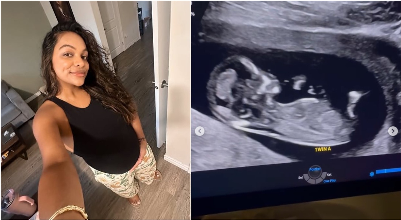
A pregnant woman discovered she was expecting twins during a routine ultrasound that simultaneously revealed a 9-centimeter ovarian cyst, according to reports from Il Messaggero. The incidental finding of the large cyst led to immediate medical intervention, which the patient later credited with saving her life.
The patient, whose identity was shared via the report, stated that the timing of the pregnancy scan was critical. She noted that without the discovery of the twins and the subsequent imaging, the cyst may have gone undetected until it caused a life-threatening complication. “Without them, I would have died,” she said, referring to the twins whose presence necessitated the ultrasound.
Ovarian cysts are fluid-filled sacs or pockets in an ovary or on its surface. While many are benign and resolve on their own, cysts reaching 9 centimeters are considered large and increase the risk of ovarian torsion—a condition where the ovary twists around its blood supply—or rupture, both of which can require emergency surgery.
How was the ovarian cyst discovered during pregnancy?
The cyst was identified through an obstetric ultrasound, a standard procedure used to monitor fetal development. According to the report, the scan was initially intended to check on the health of the fetuses. Instead, the imaging captured a significant mass on the ovary measuring 9 centimeters.
Medical professionals use ultrasound to visualize the pelvic region during pregnancy. Because the uterus expands and displaces other organs, anomalies in the ovaries can sometimes be more apparent or, conversely, masked by the pregnancy. In this case, the specific placement and size of the cyst made it visible to the sonographer.
According to the Mayo Clinic, most ovarian cysts are harmless, but larger cysts are more likely to cause symptoms such as pelvic pain or a feeling of fullness in the abdomen.
What are the risks of a 9-centimeter ovarian cyst?
A cyst of this size poses several clinical risks, particularly during pregnancy when the abdominal cavity is under increased pressure. The primary concern for a 9-centimeter mass is ovarian torsion. This occurs when the weight of the cyst causes the ovary to rotate, cutting off the blood supply to the ovarian tissue, which can lead to necrosis if not treated immediately.

Another risk is rupture. A ruptured cyst can cause internal bleeding and acute abdominal pain, potentially mimicking the symptoms of appendicitis or other surgical emergencies. During pregnancy, these symptoms can sometimes be confused with normal prenatal changes, making early detection via imaging vital.
The NHS notes that while many cysts disappear without treatment, those that are large or cause pain may require surgical removal, typically via a laparoscopic procedure or a laparotomy, depending on the patient’s condition and the stage of pregnancy.
Managing ovarian cysts during a twin pregnancy
The presence of twins increases the rate of uterine expansion compared to a singleton pregnancy, which can further compress the pelvic organs. This puts additional pressure on a pre-existing ovarian cyst, increasing the likelihood of torsion as the pregnancy progresses.
Medical teams must balance the risk of the cyst against the risks of surgical intervention during pregnancy. For very large cysts, surgeons may opt for a “watch and wait” approach with frequent monitoring, or they may perform surgery in the second trimester, which is generally considered the safest window for non-obstetric surgery during gestation.
The patient in this case emphasized that the incidental nature of the discovery—finding the cyst while looking for the twins—was the deciding factor in her survival, as she had not experienced the typical warning signs that would have prompted a separate gynecological exam.
When should pregnant women seek additional screening?
While routine ultrasounds often catch these issues, health providers suggest that pregnant women report specific “red flag” symptoms immediately. These include sudden, severe pelvic or abdominal pain, nausea accompanied by vomiting, or a rapid increase in abdominal girth that is not consistent with fetal growth.
Because the patient in the report was asymptomatic until the scan, her case highlights the importance of the standard prenatal imaging schedule. Regular check-ups allow physicians to monitor not only the fetuses but also the maternal health markers and pelvic anatomy.
For more information on prenatal care and the management of pregnancy complications, patients are encouraged to consult the American College of Obstetricians and Gynecologists (ACOG) or their local health authority.
The patient continues to recover and monitor her pregnancy. Further updates on her health and the delivery of the twins have not been released.
Do you have experience with incidental findings during prenatal scans? Share your thoughts or questions in the comments below.