Unlocking Yellow Fever’s Secrets: New High-Resolution Images Pave the Way for Next-generation Vaccines
Yellow fever, a potentially deadly mosquito-borne viral infection, continues to threaten public health across Africa and South America. For decades, scientists have strived to understand the intricacies of this virus, hindering the development of targeted treatments. Now, groundbreaking research from the University of Queensland (UQ) has delivered a major breakthrough: the first detailed, high-resolution images of the yellow fever virus (YFV), revealing crucial differences between the protective vaccine strain and its virulent counterparts. This isn’t just a visual advancement; it’s a pivotal step towards designing more effective vaccines and potentially,antiviral therapies. But what exactly do these images reveal, and how will they impact the fight against yellow fever and related viruses?
A New View of a Long-Standing Threat
Yellow fever virus, transmitted by Aedes and Haemagogus mosquitoes, attacks the liver and can lead to severe illness, including jaundice (so the name “yellow” fever), hemorrhage, and organ failure. While a highly effective vaccine exists, understanding the virus’s structure is paramount for continuous improvement and preparedness against evolving strains.
Researchers, led by Dr. Summa Bibby of UQ’s School of Chemistry and Molecular Bioscience, have achieved what was previously unattainable: a complete 3D model of a fully mature YFV particle at near-atomic resolution. This feat was accomplished using a clever technique. Rather of directly imaging the dangerous virus, they leveraged a pre-existing, harmless “platform” - the Binjari virus, also developed at UQ. By combining the structural genes of yellow fever with the Binjari virus’s safe backbone, they created virus particles suitable for examination under a cryo-electron microscope.
“We’ve been studying yellow fever for many decades, but this is the first time we’ve been able to visualize a complete, mature virus particle in such incredible detail,” explains Dr. Bibby. The results, published in Nature Communications [https://www.nature.com/articles/s41467-023-43668-x], are already reshaping our understanding of the virus.
the Surface Tells the Story: Vaccine Strain vs. Virulent Strain
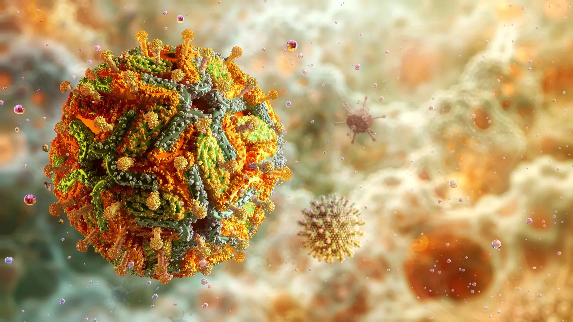
The high-resolution images revealed a striking difference between the YFV-17D vaccine strain and the strains responsible for causing illness. The vaccine strain presented a remarkably smooth and stable surface. In contrast, the virulent strains exhibited a noticeably uneven, textured exterior.
But this isn’t merely an aesthetic difference. This surface structure directly impacts how the immune system recognizes and responds to the virus.
“The bumpier, irregular surface of the virulent strains exposes parts of the virus that are normally hidden,” Dr. Bibby clarifies. “This allows certain antibodies to attach more easily.” Conversely, the smooth surface of the vaccine strain keeps these vulnerable regions covered, making it harder for antibodies to bind. This difference in antibody accessibility is a key factor in the vaccine’s safety and efficacy.
Why This Matters: Implications for Vaccine Development and Global health
Yellow fever remains a critically important public health concern, with outbreaks occurring regularly in regions with limited access to vaccination. According to the World Health Organization (WHO), in 2022, 18 countries reported cases of yellow fever, resulting in over 900 confirmed cases and 30,000 suspected cases [https://www.who.int/news-room/fact-sheets/detail/yellow-fever]. Currently, vaccination is the primary preventative measure, as no approved antiviral treatments exist.
Professor Daniel watterson emphasizes the far-reaching implications of this research. “The yellow fever vaccine remains remarkably effective against modern strains, and seeing the virus in such fine detail lets us better understand why the vaccine strain behaves the way it does.”
This detailed understanding allows scientists to pinpoint the specific structural features responsible for the vaccine’s safety and effectiveness. More importantly, the insights gained from studying yellow fever could inform the development of improved vaccines for related viruses, including:
* Dengue Fever: A mosquito-borne viral infection affecting millions globally.
* Zika Virus: Known for causing birth defects, particularly microcephaly.
* West nile Virus: A widespread virus that can cause neurological illness.
These viruses belong to the Flaviviridae family, sharing significant structural similarities with yellow fever. By applying the knowledge gained from this research, scientists can potentially accelerate the development of more effective vaccines and antiviral therapies for these debilitating diseases. Recent studies (within the last 6 months) have shown promising results in utilizing structural biology to design broadly