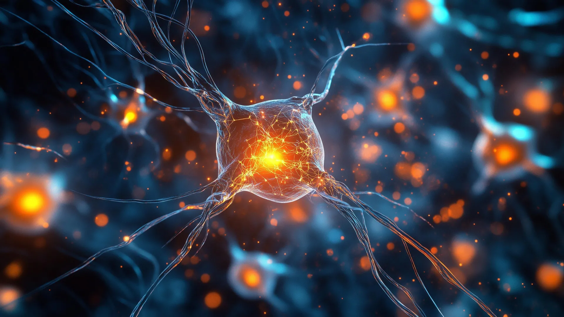
Breakthrough in Parkinson’s Research: New Technique Visualizes Disease at its Earliest Stages
For over a century, Parkinson’s disease has remained a formidable neurological challenge. Now, a groundbreaking new technique is offering an unprecedented glimpse into the very beginnings of this debilitating condition, potentially revolutionizing diagnosis, treatment, and our basic understanding of its progression. Researchers at Cambridge, Polytechnique Montréal, and The francis Crick Institute have developed a method capable of visualizing the microscopic protein clusters believed to be the true drivers of Parkinson’s – a feat previously considered impossible.
The Growing Burden of Parkinson’s Disease
Parkinson’s disease affects over 166,000 individuals in the UK alone, with a projected global prevalence of 25 million by 2050. while current medications can manage symptoms like tremors and rigidity, they offer no cure and fail to address the underlying disease process.This underscores the urgent need for innovative research focused on early detection and disease-modifying therapies.
Beyond Lewy Bodies: Focusing on the Root Cause
Traditionally, Parkinson’s diagnosis has relied on identifying Lewy bodies – large, abnormal protein deposits found in the brains of affected individuals. However, mounting evidence suggests that these Lewy bodies are a result of the disease, not its initial cause. The real culprits are thought to be smaller, more elusive structures called oligomers – early-stage protein clusters that disrupt brain cell function.
“Lewy bodies are the hallmark of Parkinson’s, but they essentially tell you where the disease has been, not where it is right now,” explains Professor Steven Lee of Cambridge’s Yusuf Hamied Department of Chemistry, a co-leader of the study. “Identifying and studying these early oligomers is crucial to understanding how Parkinson’s develops and, ultimately, how to stop it.”
ASA-PD: Illuminating the Invisible
The research team has overcome the long-standing challenge of visualizing these nanometer-sized oligomers with a novel technique called ASA-PD (Advanced Sensing of Aggregates for Parkinson’s Disease). This ultra-sensitive fluorescence microscopy method dramatically enhances the faint signal emitted by these tiny clusters while together minimizing background noise.The result? scientists can now clearly observe individual alpha-synuclein clusters – the protein implicated in Parkinson’s – with unprecedented clarity.
As Dr.Rebecca Andrews, co-first author of the study, aptly describes it, “This is the first time we’ve been able to look at oligomers directly in human brain tissue at this scale: it’s like being able to see stars in broad daylight.” This breakthrough opens entirely new avenues for Parkinson’s research.
Key Findings: A Clear Distinction Between Healthy and Diseased Brains
By applying ASA-PD to post-mortem brain tissue from both Parkinson’s patients and healthy individuals, the researchers uncovered significant differences. While oligomers were present in both groups, those with Parkinson’s exhibited:
* Larger Cluster Size: Oligomers in Parkinson’s brains were substantially larger than those found in healthy brains.
* Increased Brightness: The oligomers in Parkinson’s brains emitted a stronger fluorescent signal, indicating a higher degree of protein aggregation.
* Greater Abundance: Parkinson’s brains contained a far greater number of these damaging protein clusters.
Furthermore, the team identified a unique subset of oligomers exclusively found in Parkinson’s patients, potentially representing the earliest detectable biomarkers of the disease – appearing years before the onset of motor symptoms.
Implications for Future Research and Treatment
this research isn’t just about visualization; it’s about unlocking new therapeutic possibilities. Professor Lucien Weiss of Polytechnique Montréal emphasizes the potential for targeted therapies: “Oligomers have been the needle in the haystack, but now that we know where those needles are, it could help us target specific cell types in certain regions of the brain.”
the implications extend beyond Parkinson’s. Professor Sonia Gandhi of The Francis Crick Institute notes the potential for applying similar technologies to othre neurodegenerative diseases like Alzheimer’s and huntington’s.”The only real way to understand what is happening in human disease is to study the human brain directly,” she explains. “We hope that breaking through this technological barrier will allow us to understand why, where and how protein clusters form and how this changes the brain surroundings and leads to disease.”
A Collaborative Effort Fueled by Dedicated Support
This landmark research was made possible through the generous support of Aligning Science Across Parkinson’s (ASAP), the Michael J. Fox Foundation, and the Medical Research Council (MRC). The researchers also express deep gratitude to the patients, families, and caregivers who donated brain tissue, enabling this critical advancement in our understanding of Parkinson’s disease.
source: Andrews, R. et al. (2
Worth a look