Analyzing genetic data from nearly 140,000 in vitro fertilization embryos, researchers have identified specific maternal genetic variants tied to chromosomal abnormalities that drive pregnancy loss, offering new clarity on the molecular pathways influencing human reproductive success and fertility care.
Mapping the Genetic Landscape of Pregnancy Loss With IVF Embryo Data
Pregnancy loss remains widespread globally, with approximately 15 percent of known pregnancies ending in miscarriage, though true figures are likely higher due to losses occurring before detection. To uncover the underpinnings of this widespread public health challenge, researchers examined genetic data drawn from 139,416 human embryos. The investigation focused on chromosomal abnormalities, known as aneuploidies, which represent one of the most frequent biological drivers of pregnancy loss.
Roughly half of all known miscarriages occurring in the first or second trimester involve fetuses carrying an incorrect number of chromosomes, either too many or too few. While maternal age has long been established as a primary risk factor for these meiotic errors, our understanding of the broader genetic context has historically been hindered by a lack of large-scale data. Addressing this gap required analyzing an unprecedented volume of genetic material from tens of thousands of biological parents and their pre-implantation embryos.
The study utilized clinical data derived from pre-implantation genetic testing, examining 139,416 embryos contributed by 22,850 sets of biological parents. Within that massive sample, researchers identified 92,485 aneuploid chromosomes spread across 41,480 different embryos. The findings establish a clear relationship between aneuploidy and the exchange of DNA between chromosomes during female meiosis and egg formation, isolating common maternal genetic variants linked directly to meiotic errors.
Specific Meiotic Genes Linked to Chromosome Cohesion and Recombination
The strength of the findings stems directly from the vast scale of the dataset. As senior author Rajiv McCoy, a computational biologist at Johns Hopkins University, explained, reproductive success is closely tied to evolutionary survival, meaning natural selection restricts most common genetic differences to those with very small individual effects.
“This is a trait closely related to survival and reproductive success, so evolution will only allow genetic differences with small effects to be common in the population. You need large samples to be able to detect these small effects.”
Rajiv McCoy, computational biologist at Johns Hopkins University
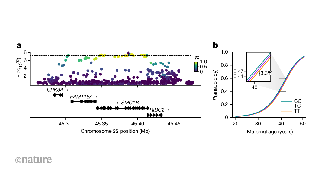
That large scale and high resolution enabled scientists to uncover several well-characterized associations between a mother’s DNA and her risk of producing embryos that fail to survive. The strongest association surfaced in genes influencing how chromosomes pair, recombine, and aggregate during meiosis in egg cell lines. Specifically, a variant of the gene SMC1B—which encodes a protein responsible for holding chromosomes together during meiosis—was associated with reduced crossover counts and an increased rate of maternal meiotic aneuploidy.

The analysis also highlighted associations with several other genes dedicated to crossover recombination, including C14orf39, CCNB1IP1, and RNF212. Commenting on these results, McCoy noted that the genetic factors emerging from the human data align precisely with mechanisms that experimental biologists have observed over decades in model organisms like mice and worms, where they play a critical role in recombination and chromosome cohesion.
“This finding is especially compelling, because the genes that emerged from our study in humans are exactly the ones that experimental biologists have detailed over decades as critical for recombination and chromosome cohesion in model organisms like mice and worms.”
Rajiv McCoy, computational biologist at Johns Hopkins University
Clinical Implications, Study Scope, and Methodological Limitations
Female meiosis begins during fetal development, when chromosomes pair and recombine before pausing for years until resuming later in life for ovulation and fertilization. Inherited differences in these processes can lead to chromosomes separating too readily during the interim, setting the stage for aneuploidy when meiosis resumes. Despite these molecular insights, experts emphasize that predicting individual risk for pregnancy loss will remain complex, as maternal age and environmental exposures continue to exert powerful influences.

Independent evaluations of the work note that while the study’s sample size, statistical power, and computational methods are robust—offering a solid framework that connects advanced genetics to human embryology—inherent design limitations must be considered. The research cohort was drawn from a specific population of infertile women undergoing IVF with pre-implantation genetic testing for aneuploidies, meaning the findings do not directly represent the general fertile population or all women experiencing spontaneous miscarriages.
Furthermore, because the genetic data relied on trophectoderm biopsies representing a small fraction of cells from a blastocyst, potential issues such as cellular mosaicism and placental representation versus inner cell mass must be kept in view. While the findings establish foundational knowledge for future reproductive genetics, drug development, and precision medicine targets, current clinical applicability remains limited because individual genetic variants carry a very low causal contribution compared to the overriding impact of maternal age.
Worth a look