
Researchers have developed machine-learning models using sleep brain wave recordings to estimate biological brain age and assess dementia risk. Published in JAMA Network Open, studies show that an older estimated brain age relative to chronological age significantly increases dementia likelihood, while metabolic health and sleep features offer new windows into cognitive aging.
A novel machine-learning approach examining brain activity during sleep is transforming how researchers identify individuals at an elevated risk of developing dementia. Led by scientists at UC San Francisco and Beth Israel Deaconess Medical Center in Boston, the system estimates a person’s brain age
by analyzing electrical signals collected via electroencephalography during the night.
Machine-Learning Models Estimate Brain Age From Sleep Signals
The research team built their predictive model by combining 13 microscopic features found within EEG brain wave recordings. They applied this algorithm to data gathered from approximately 7,000 participants across five separate studies, tracking individuals aged 40 to 94 who showed no signs of dementia at baseline. Over monitoring periods lasting from 3.5 to 17 years, roughly 1,000 participants developed dementia.
The analysis revealed that dementia risk climbed when a participant’s estimated brain age exceeded their actual chronological age. For every 10-year discrepancy where the brain appeared older than the person’s real age, the likelihood of developing dementia rose by nearly 40%. Conversely, individuals whose estimated brain age stayed younger than their chronological age faced a reduced risk.
Traditional sleep assessments—such as total sleep time, time spent in specific sleep stages, and sleep efficiency—failed to show a meaningful association with dementia risk in previous pooled analyses. By contrast, the detailed microscopic patterns in sleeping brain waves captured by the model provided crucial prognostic information.
Specific Brain Wave Patterns Connected to Memory and Risk
Several EEG patterns utilized in the brain age calculations are already known to support memory and cognitive preservation. These include delta waves, the slow electrical patterns characteristic of deep sleep, and sleep spindles, which are brief bursts of rapid brain activity that help the brain store and strengthen memories.
A particularly notable discovery involved large, sudden spikes in EEG signals known as kurtosis, which researchers linked to a lower risk of developing dementia. This heightened risk connection between an older estimated brain age and cognitive decline remained statistically significant even after adjusting for smoking, education, body mass index, physical activity, genetic risk factors, and co-occurring medical conditions.
Because EEG data can be acquired without invasive procedures, investigators suggest that wearable technologies might eventually record these critical brain signals during sleep outside traditional clinical settings. Researchers note that altering sleep health and managing body weight or exercise routines to reduce conditions like sleep apnea could influence biological brain aging, though experts emphasize there is no simple fix.
“Better body management, such as lowering body mass index and increasing exercise to reduce the likelihood of apnea, may have an impact,” said first author Haoqi Sun, PhD, assistant professor of neurology at Beth Israel Deaconess Medical Center, who developed the model with two co-authors. “But there’s no magic pill to improve brain health.”
Haoqi Sun, PhD, assistant professor of neurology at Beth Israel Deaconess Medical Center
Metabolic Syndrome Accelerates the Brain-Age Gap
Parallel research indicates that systemic health conditions can similarly drive accelerated brain aging. Affecting roughly one in four adults worldwide, metabolic syndrome is diagnosed when an individual presents with at least three of five specific risk factors: high blood sugar, high blood pressure, excess abdominal fat, high triglycerides, and low HDL cholesterol.
To study how these combined conditions impact the brain, researchers analyzed data from 27,375 adults aged 40 to 70 enrolled in the UK Biobank, utilizing artificial intelligence and more than 1,000 brain MRI imaging markers to train a machine-learning model.

The findings demonstrated a direct dose-response relationship between metabolic syndrome components and an older-looking brain. Individuals with three metabolic syndrome components had brains that looked roughly one year older than their chronological age. That gap widened to 1.7 years for those with four components and reached 2.3 years among participants presenting with all five components.
Blood sample analysis in the UK Biobank cohort highlighted three biological markers explaining part of this accelerated aging: GlycA as an indicator of chronic inflammation, the ApoB-to-ApoA1 ratio associated with blood vessel narrowing, and shifts in fatty acid levels that alter cellular structure and function.
Advanced Brain Aging in Parkinson’s Disease and Cognitive Impairment
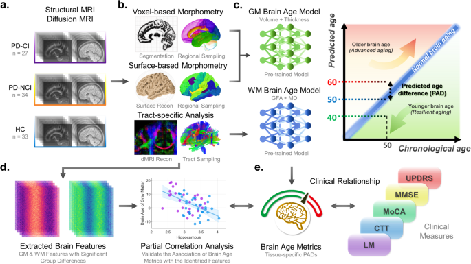
Brain-age disparities are also prominent in neurodegenerative movement disorders. A comparative study evaluated brain-age differences—known as brain-age paradigm (PAD) measures—across patients with Parkinson’s disease with cognitive impairment, Parkinson’s disease without cognitive impairment, and healthy control participants.
The study found that gray matter brain-age differences were significantly more advanced in patients with Parkinson’s disease and cognitive impairment compared to healthy controls and non-impaired patients. White matter PAD measures showed a similar pattern of accelerated aging, with statistical adjustments accounting for age, sex, and education levels.
Subgroup analyses further indicated that white matter aging measures were particularly susceptible to the combined impacts of both motor-specific symptom severity and cognitive impairment, highlighting how diverse physiological pressures converge to alter the biological trajectory of the aging brain.
Related reading