The multi-center research was conducted in accordance with the Declaration of Helsinki and secured approval from the Ethics Commission of the Medical Faculty of the University of Duisburg-Essen, referencing approval number 17-7754_2-BO. Ethics commissions at three external sites also granted approval or waived the need for additional approval due to the retrospective design, including the University of Würzburg, Centre Eugène Marquis in Rennes, and Münster under ethics approval 2018-638-f-S.
Clinical Study Design and Multicenter Patient Cohorts
The investigation gathered HCC patients treated across four distinct sites. Specifically, 75 patients treated between January 2007 and April 2015 from the University Hospital Essen in Germany formed the training cohort. Overall, half of the patients underwent liver biopsy for diagnosis, while the remaining individuals were diagnosed using imaging alone. Treatment indications for most patients included unresectability, size as a treatment alternative to transarterial chemoembolization (TACE), and 10% serving as a bridge to liver transplantation.
Inclusion Criteria and Microsphere Administration
Inclusion criteria specified that patients received RE exclusively to the right liver lobe for unresectable unilateral HCC, accompanied by computed tomography (CT) or magnetic resonance imaging (MRI) of the liver performed a maximum of six weeks prior to RE alongside at least one follow-up imaging session at least one month afterward. Exclusion criteria ruled out patients who had previous segmentectomy or surgical liver resection, as well as those who underwent a previous transarterial chemoembolization procedure within 12 months prior to RE.
Patients in the training cohort from Essen and the validation cohort from Rennes received treatment with glass 90Y microspheres. Meanwhile, the cohorts from Münster and Würzburg were treated using resin 90Y microspheres.
Volumetric Analysis and Imaging Protocols
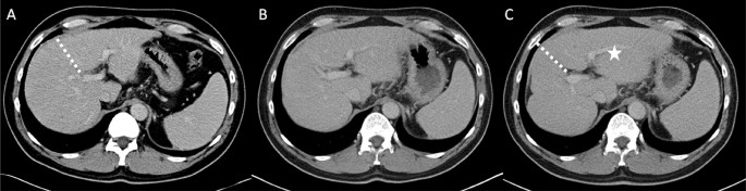
Volumetric analysis utilized baseline CT or MRI scans alongside corresponding follow-up scans captured in the portal phase. Volumetry before and after treatment was executed by an identical radiologist or nuclear medicine physician who possessed a minimum of 5 years of experience. Three distinct volumes were measured in milliliters: the left lobe, the right lobe, and the total liver volume. From these measurements, the future liver remnant (FLR) was computed as the ratio of the left lobe volume to the total volume expressed as a percentage. Additionally, clinicians measured the total splenic volume at baseline.
In the training cohort, imaging scans were scheduled at 3, 6, and 9 months following RE, though 14 patients missed scans at the 9-month mark. For these individuals, the liver volume at 9 months was estimated by calculating the mean difference between the 6-month and 9-month follow-up values across all other patients and adding this difference to the individual’s 6-month follow-up value. Imaging scans within the validation cohorts occurred at varying follow-up time points, such as 4 or 7 months, with up to three follow-up measurements available per patient. Baseline characteristics, including laboratory results, were retrieved via chart review.
Clinical Context of Radioembolization in Liver Malignancies
Intraarterial injection of the radioactive isotope 90Y delivers beta radiation with a mean decay energy of 0.94 MeV, causing cellular breakdown and tumor necrosis at the target site. The isotope maintains a half-life of approximately 64 hours with a tissue penetration depth of roughly 1 centimeter, which limits radiation exposure to the surrounding parenchyma.
Historically, hepatocellular carcinoma presented treatment challenges because systemic chemotherapy demonstrated poor response rates and external-beam radiation caused significant damage to radiosensitive liver tissue. Subsequent evaluations indicated that locoregional outcomes for 90Y radioembolization in localized disease matched or exceeded those of alternative treatments such as transarterial chemoembolization or ablation.
Keep reading