
For many women, a sudden change in menstrual patterns or unexpected spotting after menopause is a cause for immediate concern. While these symptoms can stem from various benign conditions, they are often the first warning signs of endometrial cancer—a malignancy that begins in the inner lining of the uterus. As the most common gynecologic malignancy in developed countries, understanding this disease is not just a matter of clinical interest but a necessity for public health.
Endometrial cancer, frequently referred to as uterine cancer, originates in the endometrium, the mucous membrane that lines the inside of the uterus. This tissue is remarkably dynamic. it is essential for reproduction, providing the site where a fertilized egg implants and it regenerates approximately 400 times throughout the years a person menstruates. When the growth of these cells becomes uncontrolled, it can lead to a malignancy that requires a precise, multi-modal approach to treatment.
In recent years, the medical community has shifted away from a “one size fits all” approach to uterine cancers. We are moving toward a sophisticated era of personalized medicine, where the molecular blueprint of a tumor dictates the treatment plan more than the visual appearance of the cells under a microscope. This evolution in diagnosis—combining traditional staging with molecular subtyping—is significantly improving how we predict patient outcomes and select the most effective therapies.
As a physician and health journalist, I have seen how the complexity of oncology can overwhelm patients. The goal of this review is to demystify the current landscape of endometrial cancer, from the biological risk factors that drive its rise to the cutting-edge molecular classifications that are redefining survival rates.
Understanding the Endometrium and the Path to Malignancy
To understand the cancer, one must first understand the organ. The endometrium is a specialized mucous membrane designed to support a pregnancy. Under the influence of hormones, it thickens and sheds during the menstrual cycle. However, when the balance of these hormones is disrupted—particularly when estrogen is unopposed by progesterone—the lining can become excessively thick.
This thickening is known as endometrial hyperplasia. While hyperplasia is a treatable condition, it is a critical clinical marker because it can cause abnormal menstrual bleeding and, in some cases, serve as a precursor to endometrial cancer. The transition from hyperplasia to malignancy often involves a series of genetic mutations that allow cells to grow and divide without the normal regulatory signals of the body.
The incidence of endometrial cancer is currently rising in developed nations. Clinical data suggests this trend is closely tied to two primary drivers: aging populations and obesity-related factors. Adipose tissue (body fat) can convert other hormones into estrogen, creating the “unopposed estrogen” environment that stimulates the endometrial lining to grow rapidly, thereby increasing the risk of cancerous mutations.
Clinical Presentation and Early Detection
The most significant advantage in treating endometrial cancer is that it often presents with a highly visible symptom: abnormal uterine bleeding. This may manifest as bleeding between periods, heavier-than-normal menstruation, or, most critically, any vaginal bleeding after menopause. Because these symptoms are often unmistakable, endometrial cancer is frequently diagnosed at an earlier stage than many other gynecologic cancers.
However, diagnosis requires a systematic approach. When abnormal bleeding is detected, clinicians typically employ a combination of pelvic examinations, transvaginal ultrasounds to measure the thickness of the endometrial lining, and endometrial biopsies. A biopsy allows pathologists to examine the tissue for hyperplasia or malignant cells, providing the definitive diagnosis needed to begin treatment.
The Role of FIGO Classification
Once cancer is confirmed, the next step is staging. The International Federation of Gynecology and Obstetrics (FIGO) provides the global gold standard for the classification of endometrial cancer. FIGO staging traditionally focuses on how far the tumor has spread—whether it is confined to the uterus, has invaded the muscle wall (myometrium), or has reached the lymph nodes and distant organs.
While FIGO staging remains essential for surgical planning, it is no longer the sole determinant of a patient’s prognosis. Modern oncology now integrates “molecular subtyping,” which looks at the genetic drivers of the tumor rather than just its size or location.
The Molecular Revolution: Beyond the Microscope
One of the most profound shifts in the management of endometrial cancer is the adoption of molecular subtyping, largely informed by research from The Cancer Genome Atlas. We now recognize that two tumors that look identical under a microscope may behave entirely differently because their genetic mutations are different.
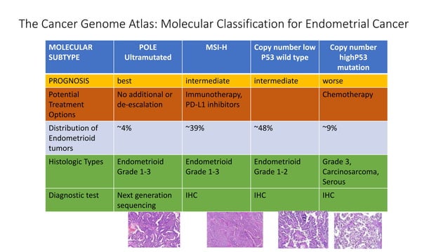
Current molecular classification generally divides endometrial cancers into four distinct groups:
- POLE-mutated: These tumors often have a very favorable prognosis, even when they appear aggressive under the microscope.
- MSI-H (Microsatellite Instability-High): These tumors have a deficiency in DNA mismatch repair, making them potentially more responsive to certain immunotherapies.
- Copy-number Low: A group with an intermediate prognosis.
- Copy-number High (Serous-like): These are generally the most aggressive tumors with a higher likelihood of spread and a poorer prognosis.
This molecular approach allows physicians to avoid “over-treating” patients with low-risk POLE-mutated tumors while intensifying treatment for those with high-risk, copy-number high malignancies. This level of precision is the cornerstone of modern personalized oncology.
Surgical Management and Adjuvant Treatment
The primary treatment for most endometrial cancers is surgical. The goal is typically the removal of the cancerous tissue and the assessment of the extent of the disease. This usually involves a total hysterectomy (removal of the uterus and cervix) and a bilateral salpingo-oophorectomy (removal of the fallopian tubes and ovaries).
In many cases, surgeons also perform lymph node sampling or a sentinel lymph node biopsy. What we have is critical for determining if the cancer has spread beyond the uterus, which directly influences whether the patient will need “adjuvant” therapy—treatment given after surgery to lower the risk of recurrence.
Adjuvant Therapy Options
Depending on the FIGO stage and the molecular subtype, adjuvant treatments may include:
- Radiation Therapy: This may be delivered as external beam radiation or brachytherapy (internal radiation) to target the pelvic area and reduce local recurrence.
- Chemotherapy: Used primarily for advanced stages or aggressive molecular subtypes (such as copy-number high) to treat systemic disease.
- Immunotherapy: A newer frontier, particularly for MSI-H tumors, where drugs are used to help the patient’s own immune system recognize and attack the cancer cells.
| Feature | Traditional (FIGO/Histologic) | Molecular Subtyping |
|---|---|---|
| Basis of Analysis | Tumor size, depth of invasion, and cell shape | Genetic mutations and DNA copy numbers |
| Primary Tool | Microscope and Surgical Staging | Genomic Sequencing (e.g., TCGA) |
| Treatment Goal | Anatomical removal of disease | Targeted therapy based on genetic drivers |
| Prognostic Value | General risk based on spread | Highly specific individual risk profiles |
Key Takeaways for Patients and Caregivers
- Early Detection is Key: Any postmenopausal bleeding or irregular uterine bleeding should be evaluated by a healthcare provider immediately.
- Obesity and Age are Factors: While not the only causes, these are significant risk factors that make regular screenings more important.
- Molecular Testing Matters: Ask your oncology team if molecular subtyping has been performed on the tumor, as this can significantly alter the treatment path.
- Multidisciplinary Care: The best outcomes typically result from a team approach involving gynecologic oncologists, pathologists, and radiation oncologists.
Looking Ahead: The Future of Uterine Cancer Care
The trajectory of endometrial cancer research is moving toward even greater precision. We are seeing an increase in the use of targeted therapies that attack specific mutations, reducing the reliance on broad-spectrum chemotherapy which often carries heavy side effects. The integration of liquid biopsies—tests that can detect cancer DNA in the blood—may soon allow us to monitor for recurrence much earlier than current imaging allows.
For those currently navigating a diagnosis, the most important takeaway is that the tools available today are far more powerful than they were even a decade ago. The combination of early detection through symptom awareness and the precision of molecular profiling is creating a new standard of care that prioritizes both survival and quality of life.
The next major milestone in this field will be the widespread standardization of molecular testing in community hospitals, ensuring that every patient, regardless of where they are treated, has access to genomic profiling. We expect updated clinical guidelines from major oncological bodies to further integrate these molecular markers into daily practice over the coming year.
Do you or a loved one have questions about the latest developments in gynecologic oncology? Share your thoughts or experiences in the comments below, and please share this guide with others who may find it helpful.