
Researchers at the University of Kentucky discovered that brain immune cells called microglia—not amyloid-beta plaques or dying neurons—drive sleep loss in an animal model of Alzheimer’s. Using a microglia-blocking drug, scientists restored more than two hours of restorative sleep nightly in mice without clearing a single plaque, pointing toward a treatment target.
Roughly a quarter to nearly half of Alzheimer’s patients experience clinically significant sleep disruptions. For years, medical science pointed the finger squarely at sticky amyloid-beta protein clumps that accumulate in the brain during the progression of the disease. A new study published in Alzheimer’s & Dementia reveals that the brain’s resident immune cells are the true culprits behind nighttime wakefulness.
Microglia and the Brain’s Inflammatory Awakening
The study investigated how brain immune cells known as microglia interact with developing amyloid plaques. While microglia normally serve a protective function by surveying the central nervous system and clearing debris, chronic overactivation turns them into agents of collateral damage.
The research team illustrated the mechanism with an analogy: imagine a small fire isolated in your kitchen. In this scenario, amyloid plaques represent the fire, while microglia act as the sprinkler system. When microglia chronically overreact to accumulating plaque burdens, they flood the brain with an inflammatory response that keeps neural networks on high alert.
“Basically, we showed that it is not the plaques themselves, or solely dysfunctional neurons, that cause sleep loss but actually microglia.”
Shannon Macauley, University of Kentucky
Shannon Macauley, an associate professor of physiology in the University of Kentucky College of Medicine, described the overactive immune response. Macauley noted that the immune cells kick off this elaborate cascade of inflammation, as if the microglia are partying all night, and keeping the brain awake.
Tracking Brainwaves With Advanced Mapping Tools
To separate Alzheimer’s-related changes from normal aging, researchers studied two cohorts of animals: mice genetically engineered to develop amyloid plaques and a control group of healthy mice. Scientists monitored the subjects at six months, when plaques first begin to emerge, and at 18 months, representing an advanced disease stage.
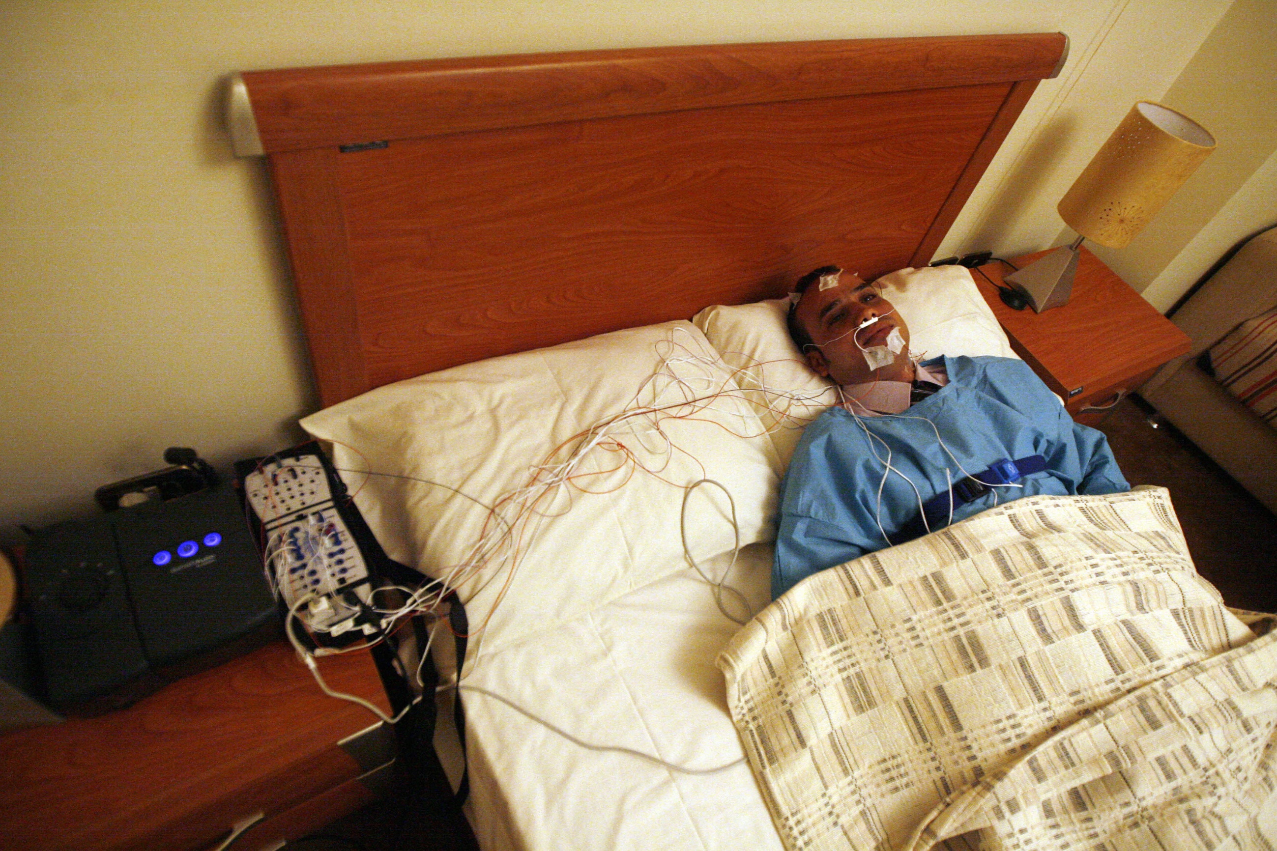
The animals wore small head-mounted devices to record electroencephalography (EEG) and electromyography (EMG) data. The EEG captured electrical fingerprints across brain networks, while the EMG tracked muscle activity. The team also utilized light-sheet microscopy, rendering brain tissue optically transparent and illuminating it with a thin laser sheet to build a comprehensive 3D digital image of both plaques and immune cells across the brain.
To evaluate electrical patterns, the team applied a mathematical algorithm known as Fitting Oscillations and One Over F (FOOOF). This technique separated periodic rhythmic sleep waves from aperiodic background noise. An elevated aperiodic slope indicated that the brain’s background activity idled at an unusually high speed, much like an engine revving while parked.
Restoring Sleep Without Clearing Plaques
To test whether microglia directly caused sleep disruption, scientists administered Pexidartinib (PLX3397). Originally developed for cancer research, the drug blocks a signaling pathway that microglia depend upon. Administering the medication for 14 days temporarily eliminated roughly 87% of the brain’s microglial population.

The intervention yielded striking results. Silencing the immune cells gave the Alzheimer’s mice an extra two hours of sleep each night. Crucially, the drug dosage left amyloid-beta plaques completely unchanged.
“That restorative sleep is super important for physical repair, learning and memory and washing out the toxins of the day.”
Shannon Macauley, University of Kentucky
When patients lose this stage of rest, Macauley explained to ScienceAlert, they lose their brain’s primary cleaning cycle, creating a feed-forward loop that may drive further damage. Furthermore, the data revealed a ceiling effect: although plaque accumulation more than doubled between six and 18 months, the resulting sleep deficit remained constant, stopping at a loss of 1.5 to 2 hours per night.
Screening Possibilities and Future Monitoring
While wiping out an entire army of brain immune cells is not a viable clinical option for humans, the findings point toward future therapies designed to calm down overactive microglia rather than removing them entirely. Researchers also noted that identifying early brain changes at the six-month mark opens new diagnostic possibilities.
The research has been published in Alzheimer’s & Dementia.
Related reading