
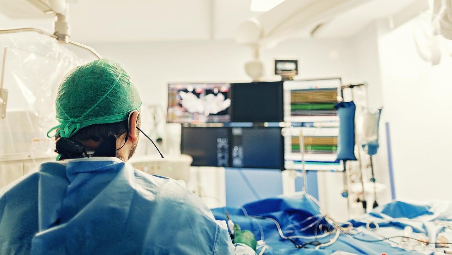
Terumo Interventional Systems (TIS) has completed the first clinical procedure in the United States using the Opuswave Dual Sensor Imaging System, according to company reports. The system provides real-time intravascular imaging to help physicians identify and treat plaque buildup in coronary arteries, a critical step in managing coronary artery disease.
The Opuswave system utilizes a combination of Optical Coherence Tomography (OCT) and Intravascular Ultrasound (IVUS) to provide a comprehensive view of the vessel wall. By integrating two distinct imaging modalities into a single catheter, the technology allows clinicians to see both the superficial layers of the artery and deeper tissue structures without switching devices during a procedure.
This development follows the broader trend of “hybrid” imaging in interventional cardiology. Traditionally, doctors chose between OCT, which offers high resolution for viewing the inner lining of the artery, and IVUS, which penetrates deeper into the vessel wall but with lower resolution. The Opuswave dual sensor system aims to eliminate the trade-off between these two methods.
How does the Opuswave Dual Sensor Imaging System work?
The Opuswave system operates by emitting both light and sound waves from a single sensor. According to Terumo Interventional Systems, the OCT component provides microscopic detail of the arterial lumen, allowing for the precise measurement of plaque thickness and the identification of thrombi (blood clots). Simultaneously, the IVUS component provides a wider view of the vessel’s external elastic lamina, which is essential for determining the correct size of a stent to be implanted.

In a standard procedure, a cardiologist inserts the imaging catheter into the coronary artery. As the device pulls back, it creates a high-definition map of the blockage. This “dual-sensor” approach reduces the time a physician spends navigating the artery, as they no longer need to perform two separate imaging passes to get a complete anatomical picture.
For patients, this efficiency can potentially lead to shorter procedure times and a reduced risk of complications associated with prolonged catheterization. The ability to accurately size stents based on both internal and external vessel diameters helps prevent “undersizing” (which can lead to restenosis) or “oversizing” (which can cause vessel trauma).
Why is dual-sensor imaging significant for coronary care?
The clinical significance of the Opuswave system lies in its ability to address “blind spots” in current imaging. According to medical literature on intravascular imaging, OCT often struggles to see through blood or deep into the vessel wall, while IVUS lacks the resolution to identify small tears or thin-cap fibroatheromas—the “vulnerable plaques” most likely to cause a heart attack.
By providing both data sets simultaneously, the system allows for “fusion imaging.” This means a cardiologist can see the high-resolution surface detail and the deep-tissue architecture in one synchronized view. This is particularly useful in complex cases, such as bifurcations (where the artery splits) or heavily calcified lesions, where precise placement of a stent is the difference between long-term success and procedural failure.
The introduction of this technology into the U.S. market expands the options for precision medicine in cardiology. It moves the field closer to a “one-stop” diagnostic approach, reducing the reliance on angiography alone, which provides only a silhouette (luminogram) of the artery rather than a cross-sectional view of the tissue.
What are the implications for U.S. healthcare providers?
The first clinical use of the Opuswave system in the U.S. signals a shift toward more integrated diagnostic tools in catheterization labs. Hospitals adopting this technology may see improvements in procedural accuracy and a potential reduction in the need for repeat interventions. Because the system streamlines the imaging process, it may also optimize the workflow within high-volume cardiac centers.
However, the adoption of such high-tech imaging depends on reimbursement policies and the willingness of providers to invest in new hardware. The value proposition for hospitals centers on the “precision” aspect—reducing the rate of stent thrombosis and improving the long-term patency of the treated vessel.

As Terumo continues to roll out the system, the medical community will be looking for comparative data to determine if dual-sensor imaging leads to statistically better patient outcomes than the sequential use of OCT and IVUS. The focus will likely remain on whether the integrated approach reduces “geographic miss”—the failure to cover the entire length of a lesion with a stent.
The next confirmed milestone for the Opuswave system will be the release of broader clinical data and adoption rates across U.S. health systems following this initial procedure. For updates on new medical device clearances and clinical trial results, readers are encouraged to monitor official FDA announcements and Terumo’s corporate filings.
Do you believe integrated imaging will become the standard of care for all stent procedures? Share your thoughts in the comments below.