
Nantes Leads Groundbreaking Cancer Imaging Trials to Improve Early Detection and Treatment
Nantes, France — In a significant leap forward for cancer diagnostics, three new clinical trials underway in Nantes are pushing the boundaries of medical imaging to enhance tumor characterization, refine diagnoses, and personalize treatment strategies. Launched under the RHU OPERANDI program, these trials aim to address critical gaps in current imaging technologies, offering hope for earlier detection and more precise therapeutic interventions for cancer patients.
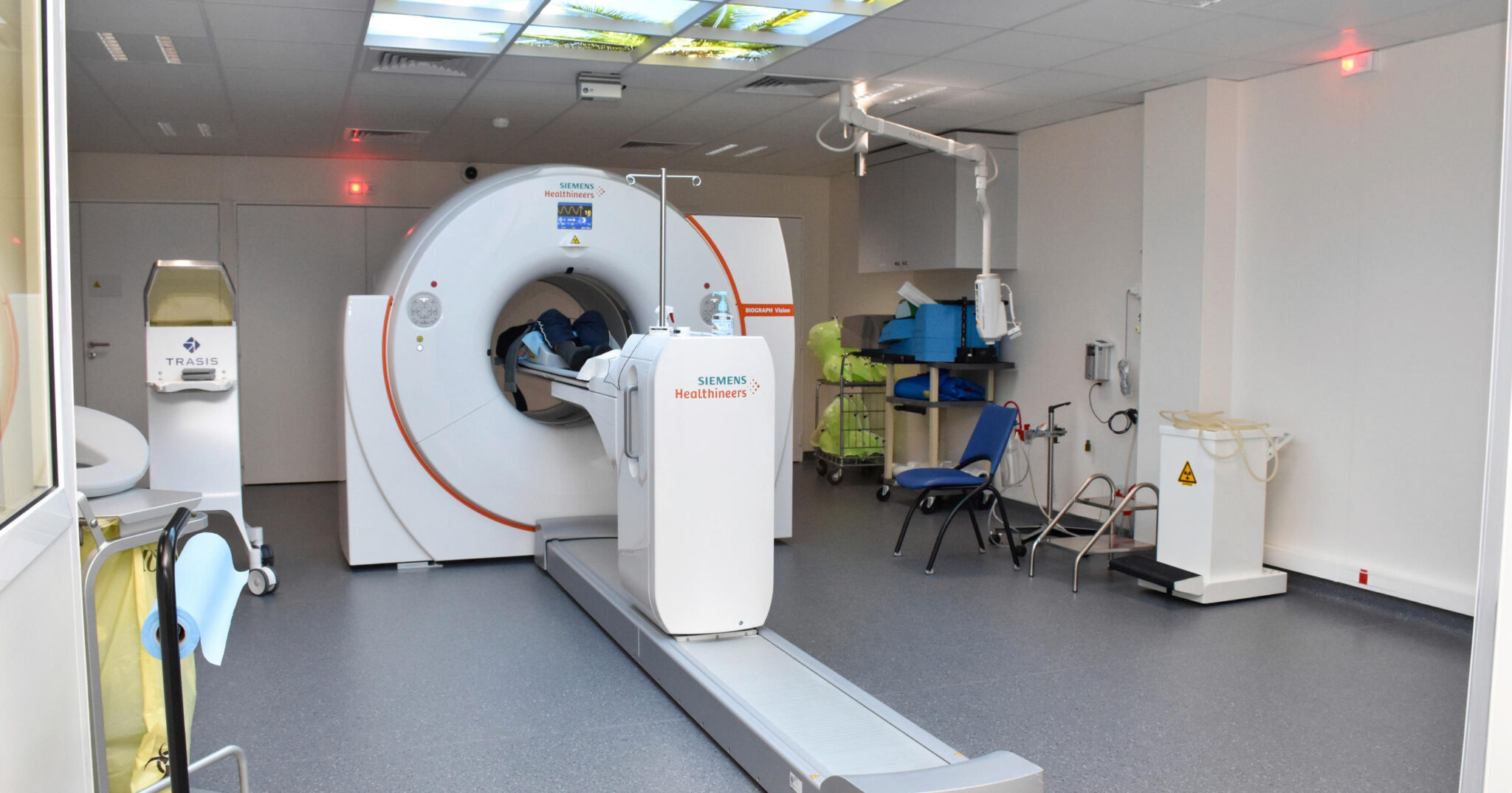
The trials, which began enrolling participants in early 2026, are part of a broader effort to integrate advanced imaging techniques with molecular and nuclear medicine. Researchers at the University Hospital of Nantes (CHU de Nantes) and the Institut de Cancérologie de l’Ouest (ICO) are collaborating with national and international partners to evaluate innovative approaches that could redefine how cancer is diagnosed and monitored.
“These trials represent a pivotal moment in oncology imaging,” said Dr. Caroline Rousseau, a nuclear medicine specialist at CHU de Nantes and a principal investigator in the OPERANDI program. “By combining cutting-edge imaging modalities with molecular insights, we are not just improving diagnostic accuracy—we are tailoring treatment pathways to the unique biology of each patient’s tumor.”
The Trials: What They Aim to Achieve
The three clinical trials focus on distinct yet complementary aspects of cancer imaging, each targeting a specific challenge in oncology diagnostics:
- Trial 1: Enhancing Tumor Characterization with Multimodal Imaging
This trial explores the use of combined positron emission tomography (PET) and magnetic resonance imaging (MRI) to improve the visualization of tumor heterogeneity. Current imaging techniques often struggle to distinguish between aggressive and indolent tumor regions, leading to suboptimal treatment decisions. By integrating PET, which highlights metabolic activity, with MRI’s high-resolution anatomical detail, researchers hope to create a more nuanced “map” of tumors, enabling clinicians to target therapies more effectively.
- Trial 2: Early Detection of Treatment Response Using Novel Radiotracers
One of the most promising aspects of the trials is the evaluation of new radiotracers—radioactive compounds used in PET scans—to detect treatment response earlier than conventional methods. Traditional imaging often requires weeks or months to reveal whether a therapy is working, delaying critical adjustments to treatment plans. The trial is testing radiotracers designed to bind to specific molecular markers associated with tumor cell death, potentially providing real-time feedback on treatment efficacy within days of administration.
- Trial 3: Personalizing Imaging Strategies for Rare and Aggressive Cancers
This trial focuses on rare and aggressive cancers, such as triple-negative breast cancer and certain types of sarcomas, where standard imaging techniques frequently fall short. Researchers are investigating the use of fluoroestradiol (FES)-PET, a specialized imaging agent that binds to estrogen receptors, to improve the detection and monitoring of hormone-sensitive tumors. The goal is to develop imaging protocols that can be tailored to the unique molecular profiles of these cancers, ultimately guiding more personalized treatment approaches.
Why These Trials Matter
Cancer remains one of the leading causes of death worldwide, with an estimated 19.3 million new cases and nearly 10 million deaths in 2020. Whereas advancements in treatment have improved survival rates for many cancers, early detection and accurate monitoring remain critical challenges. Current imaging techniques, such as computed tomography (CT) and standard PET scans, often lack the sensitivity and specificity needed to detect small tumors or distinguish between benign and malignant lesions.

The trials in Nantes aim to address these limitations by leveraging recent breakthroughs in molecular imaging and nuclear medicine. For example, the use of FES-PET in breast cancer imaging has shown promise in identifying estrogen receptor-positive tumors, which account for approximately 70% of all breast cancer cases. By improving the detection of these tumors, clinicians can better tailor hormone therapies, which are highly effective for this subtype.
“The ability to visualize tumor biology in real time is a game-changer,” said Dr. François Kraeber-Bodéré, head of nuclear medicine at CHU de Nantes and a co-investigator in the OPERANDI program. “These trials are not just about better images—they’re about transforming how we understand and treat cancer at its most fundamental level.”
The Science Behind the Innovation
The OPERANDI program, which stands for “Optimizing Personalized Radiopharmaceuticals for Enhanced Radiotherapy and Nuclear Imaging Diagnostics,” is a five-year initiative funded by the French National Research Agency (ANR) and the French National Cancer Institute (INCa). The program brings together clinicians, radiochemists, physicists, and data scientists to develop and validate next-generation imaging tools.
One of the key innovations being tested in the Nantes trials is the use of “theranostic” agents—compounds that combine diagnostic imaging with therapeutic capabilities. These agents allow clinicians to not only visualize tumors but also deliver targeted radiation directly to cancer cells. This approach is particularly promising for cancers that are resistant to conventional therapies, such as neuroendocrine tumors and certain types of prostate cancer.
Another focus of the trials is the integration of artificial intelligence (AI) into imaging workflows. AI algorithms are being trained to analyze complex imaging data, identifying subtle patterns that may be missed by the human eye. For example, AI can help differentiate between inflammation and tumor progression in post-treatment scans, reducing the risk of misdiagnosis and unnecessary interventions.
Who Stands to Benefit?
The implications of these trials extend far beyond the patients enrolled in the studies. If successful, the imaging techniques being tested could become standard practice in oncology centers worldwide, benefiting millions of cancer patients. Key groups that stand to gain include:
- Patients with Rare or Aggressive Cancers: Current imaging methods often fail to provide clear insights into rare or fast-growing tumors. The trials’ focus on molecular imaging could lead to earlier and more accurate diagnoses for these patients.
- Patients Undergoing Treatment: The ability to monitor treatment response in real time could spare patients from ineffective therapies and reduce the side effects associated with prolonged or unnecessary treatments.
- High-Risk Populations: For individuals with a genetic predisposition to cancer, such as those with BRCA mutations, advanced imaging techniques could enable earlier detection and intervention, potentially improving survival outcomes.
- Clinicians and Researchers: The data generated by these trials will provide valuable insights into tumor biology, informing future research and the development of new therapies.
Challenges and Considerations
While the potential benefits of these trials are substantial, several challenges must be addressed to ensure their success and widespread adoption:

- Cost and Accessibility: Advanced imaging techniques, particularly those involving novel radiotracers, can be expensive. Ensuring that these technologies are accessible to patients regardless of their financial or geographic circumstances will be a critical consideration.
- Regulatory Approval: New imaging agents and techniques must undergo rigorous evaluation by regulatory bodies, such as the European Medicines Agency (EMA) and the U.S. Food and Drug Administration (FDA), before they can be widely adopted. The trials in Nantes are designed to generate the data needed to support these approvals.
- Data Integration: The sheer volume of data generated by advanced imaging techniques presents challenges in terms of storage, analysis, and integration into clinical workflows. The OPERANDI program is working to develop standardized protocols for data management to ensure that imaging results are actionable and interpretable.
- Patient Participation: Clinical trials rely on the willingness of patients to participate. Ensuring that patients are fully informed about the risks and benefits of these trials is essential to their success.
What’s Next?
The three trials are expected to run for the next three to five years, with initial results anticipated in late 2027. Researchers plan to publish interim findings in peer-reviewed journals, providing the scientific community with early insights into the efficacy of the imaging techniques being tested. If the trials demonstrate significant improvements in diagnostic accuracy and treatment outcomes, the next steps will involve seeking regulatory approval and scaling up the technologies for broader use.
“Here’s just the beginning,” said Dr. Rousseau. “The ultimate goal is to develop these advanced imaging techniques a standard part of cancer care, ensuring that every patient receives the most precise and effective treatment possible.”
For patients and families affected by cancer, the trials in Nantes offer a beacon of hope. As the field of oncology imaging continues to evolve, these efforts could pave the way for a future where cancer is detected earlier, treated more effectively, and cured more often.
Key Takeaways
- Three clinical trials in Nantes are testing advanced imaging techniques to improve cancer detection and treatment. These trials are part of the RHU OPERANDI program and involve collaboration between CHU de Nantes and the Institut de Cancérologie de l’Ouest.
- The trials focus on multimodal imaging, novel radiotracers, and personalized imaging strategies for rare and aggressive cancers. These approaches aim to enhance tumor characterization, detect treatment response earlier, and tailor imaging to the unique biology of each patient’s cancer.
- Success could lead to earlier detection, more precise treatments, and improved survival outcomes for cancer patients. The trials are evaluating the integration of AI and theranostic agents, which combine diagnostic and therapeutic capabilities.
- Challenges include cost, regulatory approval, data integration, and patient participation. Addressing these challenges will be critical to ensuring the widespread adoption of these technologies.
- Initial results are expected in late 2027, with the potential for these techniques to become standard practice in oncology centers worldwide. The trials represent a significant step forward in the fight against cancer.
As these trials progress, they will undoubtedly shape the future of cancer care. For now, the medical community and patients alike are watching closely, hopeful that the innovations emerging from Nantes will bring us one step closer to a world without cancer.
What do you reckon about the potential of advanced imaging in cancer care? Share your thoughts in the comments below, and don’t forget to share this article with anyone who might locate it valuable.