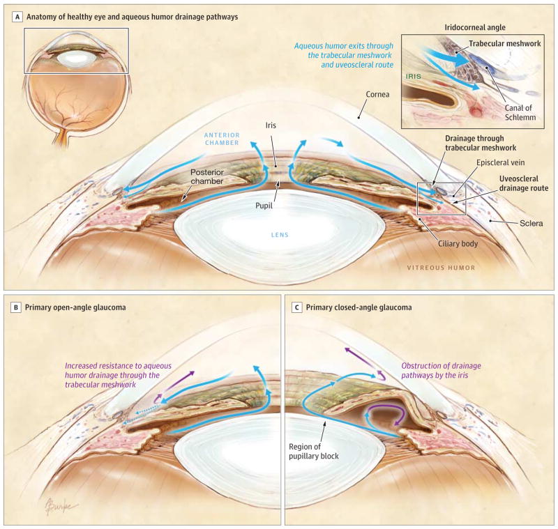
Glaucoma is a group of progressive eye conditions that damage the optic nerve, the essential pathway responsible for transmitting visual information from the retina to the brain. Often referred to as the “silent thief of sight,” this condition frequently develops without early warning signs, meaning patients may retain clear vision while irreversible nerve damage occurs. According to the World Health Organization, glaucoma is one of the leading causes of irreversible blindness globally, affecting an estimated 76 million people worldwide as of recent projections.
As a physician, I have seen firsthand how the insidious nature of this disease complicates early diagnosis. Because the damage typically begins in the peripheral vision, patients often do not notice a decline in their sight until the condition has reached an advanced stage. By the time a patient realizes their vision is compromised, the structural integrity of the optic nerve has often been significantly and permanently altered.
Understanding the Mechanics of Optic Nerve Damage
The primary mechanism driving most forms of glaucoma involves intraocular pressure (IOP). The eye contains a clear fluid, the aqueous humor, which must drain at a rate equal to its production to maintain a healthy pressure. When this drainage system becomes blocked or malfunctions, fluid accumulates, leading to increased pressure within the eye. The National Eye Institute notes that this elevated pressure can compress the optic nerve, causing the death of retinal ganglion cells that make up the nerve fibers.

While high eye pressure is a major risk factor, it is not the only cause. Normal-tension glaucoma occurs when the optic nerve is damaged even though the eye pressure remains within what is considered a “normal” range. This suggests that other factors, such as blood flow issues or structural vulnerabilities in the nerve tissue, play a critical role in the disease’s progression. Consequently, relying solely on a single pressure measurement is insufficient for a comprehensive diagnosis.
Why Early Detection Remains the Best Defense
Because vision lost to glaucoma cannot be restored, the medical community emphasizes the importance of routine screening. Comprehensive eye examinations are the only reliable way to detect the disease in its early stages. During these visits, eye care professionals perform a series of tests, including tonometry to measure eye pressure, pachymetry to measure corneal thickness, and visual field testing to map peripheral sight.

The American Academy of Ophthalmology recommends that adults with no symptoms or risk factors receive a baseline eye screening at age 40, the time when early signs of eye disease may start to appear. Individuals with higher risk factors—such as a family history of glaucoma, African, Hispanic, or Asian descent, or those with underlying conditions like diabetes—should consult their ophthalmologist about a more frequent screening schedule.
Current Treatment and Management Strategies
While there is no cure for glaucoma, modern medical interventions are highly effective at slowing or preventing further vision loss by lowering intraocular pressure. Treatment plans are highly personalized and typically follow a stepwise approach. The first line of defense usually involves daily medicated eye drops, which either reduce the production of aqueous humor or improve its drainage from the eye.
If eye drops are insufficient, laser therapy or surgical procedures may be indicated. Procedures such as trabeculectomy or the implantation of drainage devices are designed to create new pathways for fluid to exit the eye, thereby relieving pressure on the optic nerve. According to the Glaucoma Research Foundation, consistent adherence to the prescribed treatment regimen is the most significant factor in maintaining long-term visual function for patients diagnosed with the condition.
Future Directions in Glaucoma Research
Medical innovation is rapidly shifting toward neuroprotection—strategies aimed at protecting the optic nerve cells from dying, regardless of eye pressure. Researchers are currently investigating pharmacological agents and gene therapies that may stabilize the nerve tissue and prevent further degradation. While these treatments are largely in the clinical trial phase, they represent a significant shift from traditional pressure-lowering techniques toward more holistic preservation of visual function.

Patients should stay informed through their ophthalmologists regarding the availability of new diagnostic tools, such as advanced optical coherence tomography (OCT) imaging, which allows doctors to visualize the layers of the optic nerve in microscopic detail. Regular check-ups remain the most effective tool in the current clinical landscape to ensure that any progression is detected and managed promptly. For further information on local screening programs or to find a specialist, patients are encouraged to consult their national health ministry or local ophthalmological society websites.